 Movie
Movie Controller


+ Open data

- Basic information
| Entry | Database: PDB / ID: 7ox3 | ||||||
|---|---|---|---|---|---|---|---|
| Title | Fab 6D3: hIL-9 complex | ||||||
 Components Components |
| ||||||
 Keywords Keywords | CYTOKINE / IL-9 / complex | ||||||
| Function / homology |  Function and homology information Function and homology informationinterleukin-9 receptor binding / Interleukin-9 signaling / interleukin-9-mediated signaling pathway / positive regulation of interleukin-5 production / regulation of receptor signaling pathway via JAK-STAT / immunoglobulin mediated immune response / B cell proliferation / B cell differentiation / cytokine activity / growth factor activity ...interleukin-9 receptor binding / Interleukin-9 signaling / interleukin-9-mediated signaling pathway / positive regulation of interleukin-5 production / regulation of receptor signaling pathway via JAK-STAT / immunoglobulin mediated immune response / B cell proliferation / B cell differentiation / cytokine activity / growth factor activity / positive regulation of cell growth / inflammatory response / positive regulation of cell population proliferation / extracellular space / extracellular region Similarity search - Function | ||||||
| Biological species |  Homo sapiens (human) Homo sapiens (human) | ||||||
| Method |  X-RAY DIFFRACTION / SYNCHROTRON / MOLECULAR REPLACEMENT / Resolution: 1.7 Å X-RAY DIFFRACTION / SYNCHROTRON / MOLECULAR REPLACEMENT / Resolution: 1.7 Å | ||||||
 Authors Authors | De Vos, T. / Savvides, S.N. | ||||||
| Funding support |  Belgium, 1items Belgium, 1items
| ||||||
 Citation Citation | Journal: Biorxiv / Year: 2022 Title: Structural basis for the mechanism and antagonism of receptor signaling mediated by Interleukin-9 (IL-9) Authors: De Vos, T. / Godar, M. / Bick, F. / Papageorgiou, A.C. / Evangelidis, T. / Markovic, I. / Mortier, E. / Dumoutier, L. / Tripsianes, K. / Blanchetot, C. / Savvides, S.N. | ||||||
| History |
|
- Structure visualization
| Structure viewer | Molecule: MolmilJmol/JSmol |
|---|
- Downloads & links
-Download
| PDBx/mmCIF format | 7ox3.cif.gz | 232.8 KB | Display | PDBx/mmCIF format |
|---|---|---|---|---|
| PDB format | pdb7ox3.ent.gz | 184.7 KB | Display | PDB format |
| PDBx/mmJSON format | 7ox3.json.gz | Tree view | PDBx/mmJSON format | |
| Others |  Other downloads Other downloads |
-Validation report
| Arichive directory | https://data.pdbj.org/pub/pdb/validation_reports/ox/7ox3ftp://data.pdbj.org/pub/pdb/validation_reports/ox/7ox3 | HTTPS FTP |
|---|
-Related structure data
| Related structure data | 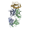 7ox1C  7ox2C 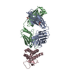 7ox4C 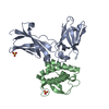 7ox5C 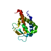 7ox6C  5nivS  6ct7S S: Starting model for refinement C: citing same article ( |
|---|---|
| Similar structure data |
-Links
 PDBj
PDBj


- Assembly
| Deposited unit | 
| ||||||||||||
|---|---|---|---|---|---|---|---|---|---|---|---|---|---|
| 1 | 
| ||||||||||||
| Unit cell |
| ||||||||||||
| Components on special symmetry positions |
|
-Components
| #1: Antibody | Mass: 22661.842 Da / Num. of mol.: 1 Source method: isolated from a genetically manipulated source Source: (gene. exp.) Homo sapiens (human) / Cell line (production host): HEK293 / Production host: Homo sapiens (human) | ||||||
|---|---|---|---|---|---|---|---|
| #2: Antibody | Mass: 23746.617 Da / Num. of mol.: 1 Source method: isolated from a genetically manipulated source Source: (gene. exp.) Homo sapiens (human) / Cell line (production host): HEK293 / Production host: Homo sapiens (human) | ||||||
| #3: Protein | Mass: 14551.010 Da / Num. of mol.: 1 Source method: isolated from a genetically manipulated source Source: (gene. exp.) Homo sapiens (human) / Gene: IL9 / Production host:  | ||||||
| #4: Chemical | ChemComp-SO4 /   Mass: 96.063 Da / Num. of mol.: 5 / Source method: obtained synthetically / Formula: SO4 Mass: 96.063 Da / Num. of mol.: 5 / Source method: obtained synthetically / Formula: SO4#5: Water | ChemComp-HOH / |  Mass: 18.015 Da / Num. of mol.: 424 / Source method: isolated from a natural source / Formula: H2O Mass: 18.015 Da / Num. of mol.: 424 / Source method: isolated from a natural source / Formula: H2OHas ligand of interest | N | Has protein modification | Y | |
-Experimental details
-Experiment
| Experiment | Method: X-RAY DIFFRACTION / Number of used crystals: 1 |
|---|
- Sample preparation
| Crystal | Density Matthews: 2.37 Å3/Da / Density % sol: 48.02 % |
|---|---|
| Crystal grow | Temperature: 293 K / Method: vapor diffusion, sitting drop / pH: 5.8 Details: 0.1M ammonium sulfate, 0.1M Tris, 16% PEG 1500, pH 5.8 |
-Data collection
| Diffraction | Mean temperature: 100 K / Serial crystal experiment: N |
|---|---|
| Diffraction source | Source: SYNCHROTRON / Site: PETRA III, EMBL c/o DESY  / Beamline: P14 (MX2) / Wavelength: 0.9763 Å / Beamline: P14 (MX2) / Wavelength: 0.9763 Å |
| Detector | Type: DECTRIS EIGER X 16M / Detector: PIXEL / Date: Aug 25, 2019 |
| Radiation | Protocol: SINGLE WAVELENGTH / Monochromatic (M) / Laue (L): M / Scattering type: x-ray |
| Radiation wavelength | Wavelength: 0.9763 Å / Relative weight: 1 |
| Reflection | Resolution: 1.7→48.64 Å / Num. obs: 63819 / % possible obs: 99.2 % / Redundancy: 13.4 % / CC1/2: 0.999 / Net I/σ(I): 21.59 |
| Reflection shell | Resolution: 1.7→1.8 Å / Num. unique obs: 10037 / CC1/2: 0.703 |
- Processing
| Software |
| ||||||||||||||||||||||||||||||||||||||||||||||||||||||||||||||||||||||||||||||||||||||||||||||||||||||||||||||||||||||||||||||||||||||||||||||||||||||||||||||||||||||||
|---|---|---|---|---|---|---|---|---|---|---|---|---|---|---|---|---|---|---|---|---|---|---|---|---|---|---|---|---|---|---|---|---|---|---|---|---|---|---|---|---|---|---|---|---|---|---|---|---|---|---|---|---|---|---|---|---|---|---|---|---|---|---|---|---|---|---|---|---|---|---|---|---|---|---|---|---|---|---|---|---|---|---|---|---|---|---|---|---|---|---|---|---|---|---|---|---|---|---|---|---|---|---|---|---|---|---|---|---|---|---|---|---|---|---|---|---|---|---|---|---|---|---|---|---|---|---|---|---|---|---|---|---|---|---|---|---|---|---|---|---|---|---|---|---|---|---|---|---|---|---|---|---|---|---|---|---|---|---|---|---|---|---|---|---|---|---|---|---|---|
| Refinement | Method to determine structure: MOLECULAR REPLACEMENT Starting model: 5NIV, 6CT7 Resolution: 1.7→48.64 Å / SU ML: 0.21 / Cross valid method: THROUGHOUT / σ(F): 1.35 / Phase error: 23.31 / Stereochemistry target values: ML
| ||||||||||||||||||||||||||||||||||||||||||||||||||||||||||||||||||||||||||||||||||||||||||||||||||||||||||||||||||||||||||||||||||||||||||||||||||||||||||||||||||||||||
| Solvent computation | Shrinkage radii: 0.9 Å / VDW probe radii: 1.11 Å / Solvent model: FLAT BULK SOLVENT MODEL | ||||||||||||||||||||||||||||||||||||||||||||||||||||||||||||||||||||||||||||||||||||||||||||||||||||||||||||||||||||||||||||||||||||||||||||||||||||||||||||||||||||||||
| Displacement parameters | Biso max: 108 Å2 / Biso mean: 40.291 Å2 / Biso min: 18.84 Å2 | ||||||||||||||||||||||||||||||||||||||||||||||||||||||||||||||||||||||||||||||||||||||||||||||||||||||||||||||||||||||||||||||||||||||||||||||||||||||||||||||||||||||||
| Refinement step | Cycle: final / Resolution: 1.7→48.64 Å
| ||||||||||||||||||||||||||||||||||||||||||||||||||||||||||||||||||||||||||||||||||||||||||||||||||||||||||||||||||||||||||||||||||||||||||||||||||||||||||||||||||||||||
| LS refinement shell | Refine-ID: X-RAY DIFFRACTION / Rfactor Rfree error: 0 / Total num. of bins used: 23
| ||||||||||||||||||||||||||||||||||||||||||||||||||||||||||||||||||||||||||||||||||||||||||||||||||||||||||||||||||||||||||||||||||||||||||||||||||||||||||||||||||||||||
| Refinement TLS params. | Method: refined / Refine-ID: X-RAY DIFFRACTION
| ||||||||||||||||||||||||||||||||||||||||||||||||||||||||||||||||||||||||||||||||||||||||||||||||||||||||||||||||||||||||||||||||||||||||||||||||||||||||||||||||||||||||
| Refinement TLS group |
|
