 Movie
Movie Controller

[English] 日本語
 Yorodumi
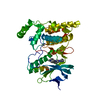
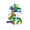
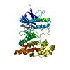
- PDB-7ore: Crystal structure of JNK3 in complex with light-activated covalen... -
+ Open data

- Basic information
| Entry | Database: PDB / ID: 7ore | ||||||
|---|---|---|---|---|---|---|---|
| Title | Crystal structure of JNK3 in complex with light-activated covalent inhibitor MR-II-249 with both non-covalent and covalent binding modes (compound 4) | ||||||
 Components Components | Mitogen-activated protein kinase 10 | ||||||
 Keywords Keywords | TRANSFERASE / kinase / MAPK / MAPK10 / light activation / covalent inhibitor / Structural Genomics / Structural Genomics Consortium / SGC | ||||||
| Function / homology |  Function and homology information Function and homology informationJUN kinase activity / Activation of the AP-1 family of transcription factors / Fc-epsilon receptor signaling pathway / MAP kinase kinase activity / response to light stimulus / mitogen-activated protein kinase / JNK cascade / JNK (c-Jun kinases) phosphorylation and activation mediated by activated human TAK1 / FCERI mediated MAPK activation / regulation of circadian rhythm ...JUN kinase activity / Activation of the AP-1 family of transcription factors / Fc-epsilon receptor signaling pathway / MAP kinase kinase activity / response to light stimulus / mitogen-activated protein kinase / JNK cascade / JNK (c-Jun kinases) phosphorylation and activation mediated by activated human TAK1 / FCERI mediated MAPK activation / regulation of circadian rhythm / cellular senescence / rhythmic process / Oxidative Stress Induced Senescence / protein phosphorylation / protein serine kinase activity / signal transduction / mitochondrion / nucleoplasm / ATP binding / nucleus / plasma membrane / cytosol / cytoplasm Similarity search - Function | ||||||
| Biological species |  Homo sapiens (human) Homo sapiens (human) | ||||||
| Method |  X-RAY DIFFRACTION / SYNCHROTRON / MOLECULAR REPLACEMENT / Resolution: 2.18 Å X-RAY DIFFRACTION / SYNCHROTRON / MOLECULAR REPLACEMENT / Resolution: 2.18 Å | ||||||
 Authors Authors | Chaikuad, A. / Reynders, M. / Trauner, D. / Knapp, S. / Structural Genomics Consortium (SGC) | ||||||
 Citation Citation | Journal: Angew.Chem.Int.Ed.Engl. / Year: 2021 Title: Controlling the Covalent Reactivity of a Kinase Inhibitor with Light. Authors: Reynders, M. / Chaikuad, A. / Berger, B.T. / Bauer, K. / Koch, P. / Laufer, S. / Knapp, S. / Trauner, D. | ||||||
| History |
|
- Structure visualization
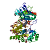
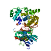
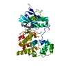
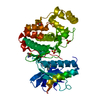
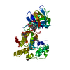
| Structure viewer | Molecule: MolmilJmol/JSmol |
|---|
- Downloads & links
-Download
| PDBx/mmCIF format | 7ore.cif.gz | 162.8 KB | Display | PDBx/mmCIF format |
|---|---|---|---|---|
| PDB format | pdb7ore.ent.gz | 128.1 KB | Display | PDB format |
| PDBx/mmJSON format | 7ore.json.gz | Tree view | PDBx/mmJSON format | |
| Others |  Other downloads Other downloads |
-Validation report
| Summary document | 7ore_validation.pdf.gz | 1.1 MB | Display | wwPDB validaton report |
|---|---|---|---|---|
| Full document | 7ore_full_validation.pdf.gz | 1.1 MB | Display | |
| Data in XML | 7ore_validation.xml.gz | 18.4 KB | Display | |
| Data in CIF | 7ore_validation.cif.gz | 24.4 KB | Display | |
| Arichive directory | https://data.pdbj.org/pub/pdb/validation_reports/or/7oreftp://data.pdbj.org/pub/pdb/validation_reports/or/7ore | HTTPS FTP |
-Related structure data
| Related structure data |  7orfC  4x21S S: Starting model for refinement C: citing same article ( |
|---|---|
| Similar structure data |


-Links
 PDBj
PDBj


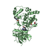
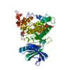
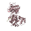
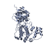
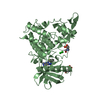
- Assembly
| Deposited unit | 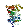
| ||||||||
|---|---|---|---|---|---|---|---|---|---|
| 1 |
| ||||||||
| Unit cell |
|
-Components
| #1: Protein | Mass: 42146.750 Da / Num. of mol.: 1 Source method: isolated from a genetically manipulated source Source: (gene. exp.) Homo sapiens (human) / Gene: MAPK10, JNK3, JNK3A, PRKM10, SAPK1B / Plasmid: pNIC28-Bsa4 / Production host:  References: UniProt: P53779, mitogen-activated protein kinase | ||||||||
|---|---|---|---|---|---|---|---|---|---|
| #2: Chemical |   Mass: 648.795 Da / Num. of mol.: 2 / Source method: obtained synthetically / Formula: C36H37FN8OS / Feature type: SUBJECT OF INVESTIGATION Mass: 648.795 Da / Num. of mol.: 2 / Source method: obtained synthetically / Formula: C36H37FN8OS / Feature type: SUBJECT OF INVESTIGATION#3: Chemical | ChemComp-EDO /   Mass: 62.068 Da / Num. of mol.: 9 / Source method: obtained synthetically / Formula: C2H6O2 Mass: 62.068 Da / Num. of mol.: 9 / Source method: obtained synthetically / Formula: C2H6O2#4: Water | ChemComp-HOH / |  Mass: 18.015 Da / Num. of mol.: 56 / Source method: isolated from a natural source / Formula: H2O Mass: 18.015 Da / Num. of mol.: 56 / Source method: isolated from a natural source / Formula: H2OHas ligand of interest | Y | Has protein modification | N | |
-Experimental details
-Experiment
| Experiment | Method: X-RAY DIFFRACTION / Number of used crystals: 1 |
|---|
- Sample preparation
| Crystal | Density Matthews: 2.39 Å3/Da / Density % sol: 48.63 % |
|---|---|
| Crystal grow | Temperature: 277.15 K / Method: vapor diffusion, sitting drop / pH: 7.8 Details: 16% medium-molecular weight PEG smears (MMW PEG Smears) and 0.1 M HEPES, pH 7.8 |
-Data collection
| Diffraction | Mean temperature: 100 K / Serial crystal experiment: N | |||||||||||||||||||||||||||
|---|---|---|---|---|---|---|---|---|---|---|---|---|---|---|---|---|---|---|---|---|---|---|---|---|---|---|---|---|
| Diffraction source | Source: SYNCHROTRON / Site: Diamond  / Beamline: I04 / Wavelength: 0.9795 Å / Beamline: I04 / Wavelength: 0.9795 Å | |||||||||||||||||||||||||||
| Detector | Type: DECTRIS PILATUS3 6M / Detector: PIXEL / Date: Jul 21, 2018 | |||||||||||||||||||||||||||
| Radiation | Protocol: SINGLE WAVELENGTH / Monochromatic (M) / Laue (L): M / Scattering type: x-ray | |||||||||||||||||||||||||||
| Radiation wavelength | Wavelength: 0.9795 Å / Relative weight: 1 | |||||||||||||||||||||||||||
| Reflection | Resolution: 2.18→29.65 Å / Num. obs: 21811 / % possible obs: 99.9 % / Redundancy: 7.1 % / CC1/2: 0.997 / Rmerge(I) obs: 0.101 / Rpim(I) all: 0.044 / Rrim(I) all: 0.118 / Net I/av σ(I): 10.6 / Net I/σ(I): 10.6 | |||||||||||||||||||||||||||
| Reflection shell | Diffraction-ID: 1
|
- Processing
| Software |
| ||||||||||||||||||||||||||||||||||||||||||||||||||||||||||||||||||||||||||||||||||||||||||||||||||||
|---|---|---|---|---|---|---|---|---|---|---|---|---|---|---|---|---|---|---|---|---|---|---|---|---|---|---|---|---|---|---|---|---|---|---|---|---|---|---|---|---|---|---|---|---|---|---|---|---|---|---|---|---|---|---|---|---|---|---|---|---|---|---|---|---|---|---|---|---|---|---|---|---|---|---|---|---|---|---|---|---|---|---|---|---|---|---|---|---|---|---|---|---|---|---|---|---|---|---|---|---|---|
| Refinement | Method to determine structure: MOLECULAR REPLACEMENT Starting model: 4x21 Resolution: 2.18→29.65 Å / Cor.coef. Fo:Fc: 0.952 / Cor.coef. Fo:Fc free: 0.915 / SU B: 16.962 / SU ML: 0.203 / SU R Cruickshank DPI: 0.2906 / Cross valid method: THROUGHOUT / σ(F): 0 / ESU R: 0.291 / ESU R Free: 0.236 / Stereochemistry target values: MAXIMUM LIKELIHOOD Details: U VALUES : WITH TLS ADDED HYDROGENS HAVE BEEN ADDED IN THE RIDING POSITIONS
| ||||||||||||||||||||||||||||||||||||||||||||||||||||||||||||||||||||||||||||||||||||||||||||||||||||
| Solvent computation | Ion probe radii: 0.8 Å / Shrinkage radii: 0.8 Å / VDW probe radii: 1.2 Å / Solvent model: MASK | ||||||||||||||||||||||||||||||||||||||||||||||||||||||||||||||||||||||||||||||||||||||||||||||||||||
| Displacement parameters | Biso max: 115.9 Å2 / Biso mean: 54.154 Å2 / Biso min: 31.22 Å2
| ||||||||||||||||||||||||||||||||||||||||||||||||||||||||||||||||||||||||||||||||||||||||||||||||||||
| Refinement step | Cycle: final / Resolution: 2.18→29.65 Å
| ||||||||||||||||||||||||||||||||||||||||||||||||||||||||||||||||||||||||||||||||||||||||||||||||||||
| Refine LS restraints |
| ||||||||||||||||||||||||||||||||||||||||||||||||||||||||||||||||||||||||||||||||||||||||||||||||||||
| LS refinement shell | Resolution: 2.18→2.236 Å / Rfactor Rfree error: 0 / Total num. of bins used: 20
| ||||||||||||||||||||||||||||||||||||||||||||||||||||||||||||||||||||||||||||||||||||||||||||||||||||
| Refinement TLS params. | Method: refined / Refine-ID: X-RAY DIFFRACTION
| ||||||||||||||||||||||||||||||||||||||||||||||||||||||||||||||||||||||||||||||||||||||||||||||||||||
| Refinement TLS group |
|
