ムービー
ムービー コントローラー


+ データを開く

- 基本情報
基本情報
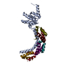
| 登録情報 | データベース: PDB / ID: 7ood | ||||||||||||
|---|---|---|---|---|---|---|---|---|---|---|---|---|---|
| タイトル | Mycoplasma pneumoniae 50S subunit of ribosomes in chloramphenicol-treated cells | ||||||||||||
 要素 要素 |
| ||||||||||||
 キーワード キーワード | RIBOSOME / In-cell cryo-electron tomography chloramphenicol-treated sub-tomogram analysis | ||||||||||||
| 機能・相同性 |  機能・相同性情報 機能・相同性情報large ribosomal subunit / transferase activity / 5S rRNA binding / ribosomal large subunit assembly / large ribosomal subunit rRNA binding / cytosolic large ribosomal subunit / cytoplasmic translation / tRNA binding / negative regulation of translation / rRNA binding ...large ribosomal subunit / transferase activity / 5S rRNA binding / ribosomal large subunit assembly / large ribosomal subunit rRNA binding / cytosolic large ribosomal subunit / cytoplasmic translation / tRNA binding / negative regulation of translation / rRNA binding / structural constituent of ribosome / ribosome / translation / ribonucleoprotein complex / response to antibiotic / mRNA binding / cytoplasm 類似検索 - 分子機能 | ||||||||||||
| 生物種 |  Mycoplasma pneumoniae (バクテリア) Mycoplasma pneumoniae (バクテリア) | ||||||||||||
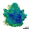
| 手法 | 電子顕微鏡法 / サブトモグラム平均法 / クライオ電子顕微鏡法 / 解像度: 3.4 Å | ||||||||||||
 データ登録者 データ登録者 | Xue, L. / Lenz, S. / Rappsilber, J. / Mahamid, J. | ||||||||||||
| 資金援助 |  ドイツ, ドイツ,  英国, 3件 英国, 3件
| ||||||||||||
 引用 引用 | ジャーナル: Nature / 年: 2022 タイトル: Visualizing translation dynamics at atomic detail inside a bacterial cell. 著者: Liang Xue / Swantje Lenz / Maria Zimmermann-Kogadeeva / Dimitry Tegunov / Patrick Cramer / Peer Bork / Juri Rappsilber / Julia Mahamid /  要旨: Translation is the fundamental process of protein synthesis and is catalysed by the ribosome in all living cells. Here we use advances in cryo-electron tomography and sub-tomogram analysis to ...Translation is the fundamental process of protein synthesis and is catalysed by the ribosome in all living cells. Here we use advances in cryo-electron tomography and sub-tomogram analysis to visualize the structural dynamics of translation inside the bacterium Mycoplasma pneumoniae. To interpret the functional states in detail, we first obtain a high-resolution in-cell average map of all translating ribosomes and build an atomic model for the M. pneumoniae ribosome that reveals distinct extensions of ribosomal proteins. Classification then resolves 13 ribosome states that differ in their conformation and composition. These recapitulate major states that were previously resolved in vitro, and reflect intermediates during active translation. On the basis of these states, we animate translation elongation inside native cells and show how antibiotics reshape the cellular translation landscapes. During translation elongation, ribosomes often assemble in defined three-dimensional arrangements to form polysomes. By mapping the intracellular organization of translating ribosomes, we show that their association into polysomes involves a local coordination mechanism that is mediated by the ribosomal protein L9. We propose that an extended conformation of L9 within polysomes mitigates collisions to facilitate translation fidelity. Our work thus demonstrates the feasibility of visualizing molecular processes at atomic detail inside cells. #1: ジャーナル: Nature / 年: 2022タイトル: Visualizing translation dynamics at atomic detail inside a bacterial cell. 著者: Liang Xue / Swantje Lenz / Maria Zimmermann-Kogadeeva / Dimitry Tegunov / Patrick Cramer / Peer Bork / Juri Rappsilber / Julia Mahamid / 要旨: Translation is the fundamental process of protein synthesis and is catalysed by the ribosome in all living cells. Here we use advances in cryo-electron tomography and sub-tomogram analysis to ...Translation is the fundamental process of protein synthesis and is catalysed by the ribosome in all living cells. Here we use advances in cryo-electron tomography and sub-tomogram analysis to visualize the structural dynamics of translation inside the bacterium Mycoplasma pneumoniae. To interpret the functional states in detail, we first obtain a high-resolution in-cell average map of all translating ribosomes and build an atomic model for the M. pneumoniae ribosome that reveals distinct extensions of ribosomal proteins. Classification then resolves 13 ribosome states that differ in their conformation and composition. These recapitulate major states that were previously resolved in vitro, and reflect intermediates during active translation. On the basis of these states, we animate translation elongation inside native cells and show how antibiotics reshape the cellular translation landscapes. During translation elongation, ribosomes often assemble in defined three-dimensional arrangements to form polysomes. By mapping the intracellular organization of translating ribosomes, we show that their association into polysomes involves a local coordination mechanism that is mediated by the ribosomal protein L9. We propose that an extended conformation of L9 within polysomes mitigates collisions to facilitate translation fidelity. Our work thus demonstrates the feasibility of visualizing molecular processes at atomic detail inside cells. #2: ジャーナル: Biorxiv / 年: 2021タイトル: Visualizing translation dynamics at atomic detail inside a bacterial cell 著者: Xue, L. / Lenz, S. / Zimmermann-Kogadeeva, M. / Tegunov, D. / Cramer, P. / Bork, P. / Rappsilber, J. / Mahamid, J. | ||||||||||||
| 履歴 |
|
- 構造の表示
構造の表示
| 構造ビューア | 分子: MolmilJmol/JSmol |
|---|
- ダウンロードとリンク
-ダウンロード
| PDBx/mmCIF形式 | 7ood.cif.gz | 2 MB | 表示 | PDBx/mmCIF形式 |
|---|---|---|---|---|
| PDB形式 | pdb7ood.ent.gz | 1.5 MB | 表示 | PDB形式 |
| PDBx/mmJSON形式 | 7ood.json.gz | ツリー表示 | PDBx/mmJSON形式 | |
| その他 |  その他のダウンロード その他のダウンロード |
-検証レポート
| 文書・要旨 | 7ood_validation.pdf.gz | 638.8 KB | 表示 | wwPDB検証レポート |
|---|---|---|---|---|
| 文書・詳細版 | 7ood_full_validation.pdf.gz | 725.3 KB | 表示 | |
| XML形式データ | 7ood_validation.xml.gz | 95.3 KB | 表示 | |
| CIF形式データ | 7ood_validation.cif.gz | 169.7 KB | 表示 | |
| アーカイブディレクトリ | https://data.pdbj.org/pub/pdb/validation_reports/oo/7oodftp://data.pdbj.org/pub/pdb/validation_reports/oo/7ood | HTTPS FTP |
-関連構造データ
| 関連構造データ |  7oocC 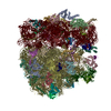 7p6zC 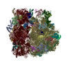 7pahC 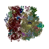 7paiC 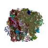 7pajC 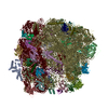 7pakC 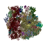 7palC 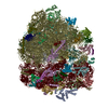 7pamC 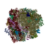 7panC 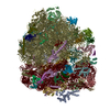 7paoC 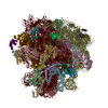 7paqC 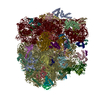 7parC 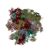 7pasC  7patC  7pauC 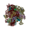 7ph9C 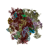 7phaC 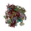 7phbC 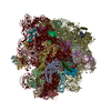 7phcC 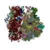 7pi8C 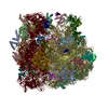 7pi9C 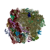 7piaC 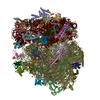 7pibC 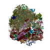 7picC 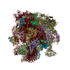 7pioC 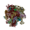 7pipC 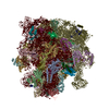 7piqC 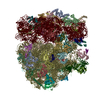 7pirC 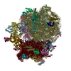 7pisC 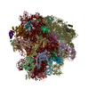 7pitC M: このデータのモデリングに利用したマップデータ C: 同じ文献を引用 ( |
|---|---|
| 類似構造データ | |
| 電子顕微鏡画像生データ | EMPIAR-10499 (タイトル: Tilt series of native M. pneumoniae cells treated with chloramphenicol Data size: 83.8 Data #1: Unaligned tilt movies of M. pneumoniae [tilt series]) EMPIAR-10731 (タイトル: Locating Macromolecular Assemblies in Cells by 2D Template Matching with cisTEMData size: 67.8 Data #1: 50S templates of Mycoplasma pneumoniae used for 2D and 3D template matching [reconstructed volumes] Data #2: Single-exposure micrographs of untilted Mycoplasma pneumoniae cells [micrographs - single frame] Data #3: Tomographic tilt series of Mycoplasma pneumoniae cells [tilt series] Data #4: Tomographic reconstructions of Mycoplasma pneumoniae cells [reconstructed volumes]) |
| 実験データセット #1 | データ参照: 10.6019/EMPIAR-10499 / データの種類: EMPIAR |


-リンク
 PDBj
PDBj


- 集合体
集合体
| 登録構造単位 | 
|
|---|---|
| 1 |
|
-要素
-RNA鎖 , 2種, 2分子 34
| #1: RNA鎖 | 分子量: 940911.500 Da / 分子数: 1 / 由来タイプ: 天然 由来: (天然) Mycoplasma pneumoniae (strain ATCC 29342 / M129) (バクテリア)株: ATCC 29342 / M129 |
|---|---|
| #2: RNA鎖 | 分子量: 34796.676 Da / 分子数: 1 / 由来タイプ: 天然 由来: (天然) Mycoplasma pneumoniae (strain ATCC 29342 / M129) (バクテリア)株: ATCC 29342 / M129 |
+50S ribosomal protein ... , 29種, 29分子 wacekimquy021osvxzdblpjntrfhg
-非ポリマー , 4種, 30分子 


| #32: 化合物 | ChemComp-K /  分子量: 39.098 Da / 分子数: 1 / 由来タイプ: 合成 / 式: K 分子量: 39.098 Da / 分子数: 1 / 由来タイプ: 合成 / 式: K | ||||
|---|---|---|---|---|---|
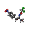
| #33: 化合物 | ChemComp-MG /  分子量: 24.305 Da / 分子数: 25 / 由来タイプ: 合成 / 式: Mg 分子量: 24.305 Da / 分子数: 25 / 由来タイプ: 合成 / 式: Mg#34: 化合物 | ChemComp-CLM / |  分子量: 323.129 Da / 分子数: 1 / 由来タイプ: 合成 / 式: C11H12Cl2N2O5 / コメント: 抗生剤*YM 分子量: 323.129 Da / 分子数: 1 / 由来タイプ: 合成 / 式: C11H12Cl2N2O5 / コメント: 抗生剤*YM#35: 化合物 |  分子量: 65.409 Da / 分子数: 3 / 由来タイプ: 合成 / 式: Zn 分子量: 65.409 Da / 分子数: 3 / 由来タイプ: 合成 / 式: Zn |
-詳細
| 研究の焦点であるリガンドがあるか | N |
|---|---|
| Has protein modification | Y |
-実験情報
-実験
| 実験 | 手法: 電子顕微鏡法 |
|---|---|
| EM実験 | 試料の集合状態: CELL / 3次元再構成法: サブトモグラム平均法 |
- 試料調製
試料調製
| 構成要素 | 名称: cryo-electron tomograms of chloramphenicol-treated Mycoplasma pneumoniae cells タイプ: RIBOSOME 詳細: ribosome sub-tomograms extracted in silico from cellular tomograms, focused refinement on 50S Entity ID: #1-#31 / 由来: NATURAL |
|---|---|
| 由来(天然) | 生物種: Mycoplasma pneumoniae (strain ATCC 29342 / M129) (バクテリア) |
| 緩衝液 | pH: 7.4 |
| 試料 | 包埋: NO / シャドウイング: NO / 染色: NO / 凍結: YES 詳細: Mycoplasma pneumoniae M129 cells grown on gold Quantifoil grids at 37 degrees Celsius before plunge freezing. |
| 試料支持 | グリッドの材料: GOLD / グリッドのタイプ: Quantifoil R2/1 |
| 急速凍結 | 装置: HOMEMADE PLUNGER / 凍結剤: ETHANE-PROPANE 詳細: ack-side blotting for 2-3 second before plunging using a manual plunger without an environmental chamber |
- 電子顕微鏡撮影
| 実験機器 |  モデル: Titan Krios / 画像提供: FEI Company |
|---|---|
| 顕微鏡 | モデル: FEI TITAN KRIOS |
| 電子銃 | 電子線源:  FIELD EMISSION GUN / 加速電圧: 300 kV / 照射モード: FLOOD BEAM FIELD EMISSION GUN / 加速電圧: 300 kV / 照射モード: FLOOD BEAM |
| 電子レンズ | モード: BRIGHT FIELD / 倍率(公称値): 81000 X / 最大 デフォーカス(公称値): 3750 nm / 最小 デフォーカス(公称値): 1500 nm / Cs: 2.7 mm |
| 試料ホルダ | 凍結剤: NITROGEN 試料ホルダーモデル: FEI TITAN KRIOS AUTOGRID HOLDER |
| 撮影 | 電子線照射量: 3.2 e/Å2 / 検出モード: COUNTING フィルム・検出器のモデル: GATAN K2 SUMMIT (4k x 4k) |
- 解析
解析
| ソフトウェア |
| ||||||||||||||||||||||||||||||
|---|---|---|---|---|---|---|---|---|---|---|---|---|---|---|---|---|---|---|---|---|---|---|---|---|---|---|---|---|---|---|---|
| EMソフトウェア |
| ||||||||||||||||||||||||||||||
| CTF補正 | タイプ: PHASE FLIPPING AND AMPLITUDE CORRECTION | ||||||||||||||||||||||||||||||
| 対称性 | 点対称性: C1 (非対称) | ||||||||||||||||||||||||||||||
| 3次元再構成 | 解像度: 3.4 Å / 解像度の算出法: FSC 0.143 CUT-OFF / 粒子像の数: 17890 詳細: Map generated by focused refinement on the 50S subunit in M 対称性のタイプ: POINT | ||||||||||||||||||||||||||||||
| EM volume selection | Num. of tomograms: 65 / Num. of volumes extracted: 17890 | ||||||||||||||||||||||||||||||
| 原子モデル構築 | プロトコル: AB INITIO MODEL / 空間: REAL | ||||||||||||||||||||||||||||||
| 原子モデル構築 | 3D fitting-ID: 1 / Source name: PDB / タイプ: experimental model
| ||||||||||||||||||||||||||||||
| 精密化 | 交差検証法: NONE 立体化学のターゲット値: GeoStd + Monomer Library + CDL v1.2 | ||||||||||||||||||||||||||||||
| 原子変位パラメータ | Biso mean: 83.32 Å2 | ||||||||||||||||||||||||||||||
| 拘束条件 |
|