 Movie
Movie Controller


+ Open data

- Basic information
| Entry | Database: PDB / ID: 7om5 | ||||||
|---|---|---|---|---|---|---|---|
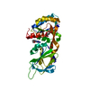
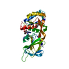
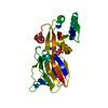
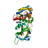
| Title | Anti-EGFR nanobody EgB4 | ||||||
 Components Components | Nanobody EgB4 | ||||||
 Keywords Keywords | IMMUNE SYSTEM / EGFR / nanobody / cancer / signaling | ||||||
| Function / homology | Immunoglobulins / Immunoglobulin-like / Sandwich / Mainly Beta Function and homology information Function and homology information | ||||||
| Biological species |  | ||||||
| Method |  X-RAY DIFFRACTION / SYNCHROTRON / MOLECULAR REPLACEMENT / Resolution: 1.48 Å X-RAY DIFFRACTION / SYNCHROTRON / MOLECULAR REPLACEMENT / Resolution: 1.48 Å | ||||||
 Authors Authors | Zeronian, M.R. / Janssen, B.J.C. | ||||||
 Citation Citation | Journal: Bmc Mol Cell Biol / Year: 2022 Title: Structural insights into the non-inhibitory mechanism of the anti-EGFR EgB4 nanobody. Authors: Zeronian, M.R. / Doulkeridou, S. / van Bergen En Henegouwen, P.M.P. / Janssen, B.J.C. | ||||||
| History |
|
- Structure visualization
| Structure viewer | Molecule: MolmilJmol/JSmol |
|---|
- Downloads & links
-Download
| PDBx/mmCIF format | 7om5.cif.gz | 68.1 KB | Display | PDBx/mmCIF format |
|---|---|---|---|---|
| PDB format | pdb7om5.ent.gz | 49.2 KB | Display | PDB format |
| PDBx/mmJSON format | 7om5.json.gz | Tree view | PDBx/mmJSON format | |
| Others |  Other downloads Other downloads |
-Validation report
| Summary document | 7om5_validation.pdf.gz | 440.2 KB | Display | wwPDB validaton report |
|---|---|---|---|---|
| Full document | 7om5_full_validation.pdf.gz | 441.3 KB | Display | |
| Data in XML | 7om5_validation.xml.gz | 13.1 KB | Display | |
| Data in CIF | 7om5_validation.cif.gz | 18.6 KB | Display | |
| Arichive directory | https://data.pdbj.org/pub/pdb/validation_reports/om/7om5ftp://data.pdbj.org/pub/pdb/validation_reports/om/7om5 | HTTPS FTP |
-Related structure data
| Related structure data |  7om4C  4krnS S: Starting model for refinement C: citing same article ( |
|---|---|
| Similar structure data |


-Links
 PDBj
PDBj


- Assembly
| Deposited unit | 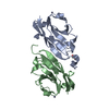
| ||||||||
|---|---|---|---|---|---|---|---|---|---|
| 1 |
| ||||||||
| Unit cell |
|
-Components
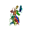
| #1: Antibody | Mass: 14195.455 Da / Num. of mol.: 2 Source method: isolated from a genetically manipulated source Source: (gene. exp.)  #2: Chemical | ChemComp-GOL / |   Mass: 92.094 Da / Num. of mol.: 1 / Source method: obtained synthetically / Formula: C3H8O3 Mass: 92.094 Da / Num. of mol.: 1 / Source method: obtained synthetically / Formula: C3H8O3#3: Chemical |   Mass: 65.409 Da / Num. of mol.: 2 / Source method: obtained synthetically / Formula: Zn Mass: 65.409 Da / Num. of mol.: 2 / Source method: obtained synthetically / Formula: Zn#4: Water | ChemComp-HOH / |  Mass: 18.015 Da / Num. of mol.: 176 / Source method: isolated from a natural source / Formula: H2O Mass: 18.015 Da / Num. of mol.: 176 / Source method: isolated from a natural source / Formula: H2OHas ligand of interest | N | Has protein modification | Y | |
|---|
-Experimental details
-Experiment
| Experiment | Method: X-RAY DIFFRACTION / Number of used crystals: 1 |
|---|
- Sample preparation
| Crystal | Density Matthews: 2.59 Å3/Da / Density % sol: 52.58 % |
|---|---|
| Crystal grow | Temperature: 293 K / Method: vapor diffusion, sitting drop / Details: 0.05 M zinc acetate and 20 % w/v PEG3350 |
-Data collection
| Diffraction | Mean temperature: 100 K / Serial crystal experiment: N |
|---|---|
| Diffraction source | Source: SYNCHROTRON / Site: Diamond  / Beamline: I24 / Wavelength: 0.9688 Å / Beamline: I24 / Wavelength: 0.9688 Å |
| Detector | Type: DECTRIS PILATUS3 6M / Detector: PIXEL / Date: Jun 27, 2019 |
| Radiation | Protocol: SINGLE WAVELENGTH / Monochromatic (M) / Laue (L): M / Scattering type: x-ray |
| Radiation wavelength | Wavelength: 0.9688 Å / Relative weight: 1 |
| Reflection | Resolution: 1.48→42.69 Å / Num. obs: 47214 / % possible obs: 98 % / Redundancy: 3.4 % / CC1/2: 0.99 / Rmerge(I) obs: 0.125 / Rpim(I) all: 0.078 / Rrim(I) all: 0.147 / Net I/σ(I): 5.4 |
| Reflection shell | Resolution: 1.48→1.5 Å / Redundancy: 3.4 % / Rmerge(I) obs: 1.681 / Mean I/σ(I) obs: 1.1 / Num. unique obs: 2189 / CC1/2: 0.277 / Rpim(I) all: 1.037 / Rrim(I) all: 1.981 / % possible all: 93 |
- Processing
| Software |
| ||||||||||||||||||||||||||||||||||||||||||||||||||||||||||||
|---|---|---|---|---|---|---|---|---|---|---|---|---|---|---|---|---|---|---|---|---|---|---|---|---|---|---|---|---|---|---|---|---|---|---|---|---|---|---|---|---|---|---|---|---|---|---|---|---|---|---|---|---|---|---|---|---|---|---|---|---|---|
| Refinement | Method to determine structure: MOLECULAR REPLACEMENT Starting model: 4KRN Resolution: 1.48→42.69 Å / Cor.coef. Fo:Fc: 0.965 / Cor.coef. Fo:Fc free: 0.958 / Cross valid method: THROUGHOUT / σ(F): 0 / ESU R: 0.067 / ESU R Free: 0.069 / Stereochemistry target values: MAXIMUM LIKELIHOOD
| ||||||||||||||||||||||||||||||||||||||||||||||||||||||||||||
| Solvent computation | Ion probe radii: 0.8 Å / Shrinkage radii: 0.8 Å / VDW probe radii: 1.2 Å / Solvent model: MASK | ||||||||||||||||||||||||||||||||||||||||||||||||||||||||||||
| Displacement parameters | Biso max: 67.05 Å2 / Biso mean: 16.232 Å2 / Biso min: 7.79 Å2
| ||||||||||||||||||||||||||||||||||||||||||||||||||||||||||||
| Refinement step | Cycle: final / Resolution: 1.48→42.69 Å
| ||||||||||||||||||||||||||||||||||||||||||||||||||||||||||||
| Refine LS restraints |
| ||||||||||||||||||||||||||||||||||||||||||||||||||||||||||||
| LS refinement shell | Resolution: 1.48→1.517 Å / Rfactor Rfree error: 0
|
