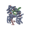
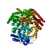
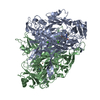
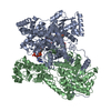
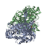
Journal: Nat Commun / Year: 2021 Title: Structure of the mini-RNA-guided endonuclease CRISPR-Cas12j3. Authors: Arturo Carabias / Anders Fuglsang / Piero Temperini / Tillmann Pape / Nicholas Sofos / Stefano Stella / Simon Erlendsson / Guillermo Montoya / Abstract: CRISPR-Cas12j is a recently identified family of miniaturized RNA-guided endonucleases from phages. These ribonucleoproteins provide a compact scaffold gathering all key activities of a genome ...CRISPR-Cas12j is a recently identified family of miniaturized RNA-guided endonucleases from phages. These ribonucleoproteins provide a compact scaffold gathering all key activities of a genome editing tool. We provide the first structural insight into the Cas12j family by determining the cryoEM structure of Cas12j3/R-loop complex after DNA cleavage. The structure reveals the machinery for PAM recognition, hybrid assembly and DNA cleavage. The crRNA-DNA hybrid is directed to the stop domain that splits the hybrid, guiding the T-strand towards the catalytic site. The conserved RuvC insertion is anchored in the stop domain and interacts along the phosphate backbone of the crRNA in the hybrid. The assembly of a hybrid longer than 12-nt activates catalysis through key functional residues in the RuvC insertion. Our findings suggest why Cas12j unleashes unspecific ssDNA degradation after activation. A site-directed mutagenesis analysis supports the DNA cutting mechanism, providing new avenues to redesign CRISPR-Cas12j nucleases for genome editing.
History
Deposition
Apr 29, 2021
Deposition site: PDBE / Processing site: PDBE
Revision 1.0
Jul 21, 2021
Provider: repository / Type: Initial release
Revision 1.0
Jul 21, 2021
Data content type: EM metadata / Data content type: EM metadata / Provider: repository / Type: Initial release
Revision 1.0
Jul 21, 2021
Data content type: Additional map / Part number: 1 / Data content type: Additional map / Provider: repository / Type: Initial release
Revision 1.0
Jul 21, 2021
Data content type: Additional map / Part number: 2 / Data content type: Additional map / Provider: repository / Type: Initial release
Revision 1.0
Jul 21, 2021
Data content type: FSC / Data content type: FSC / Provider: repository / Type: Initial release
Revision 1.0
Jul 21, 2021
Data content type: Image / Data content type: Image / Provider: repository / Type: Initial release
Revision 1.0
Jul 21, 2021
Data content type: Primary map / Data content type: Primary map / Provider: repository / Type: Initial release
Revision 1.0
Jul 21, 2021
Data content type: Additional map / Part number: 1 / Data content type: Additional map / Provider: repository / Type: Initial release
Revision 1.0
Jul 21, 2021
Data content type: Additional map / Part number: 2 / Data content type: Additional map / Provider: repository / Type: Initial release
Revision 1.0
Jul 21, 2021
Data content type: FSC / Data content type: FSC / Provider: repository / Type: Initial release
Revision 1.0
Jul 21, 2021
Data content type: Image / Data content type: Image / Provider: repository / Type: Initial release
Revision 1.0
Jul 21, 2021
Data content type: Primary map / Data content type: Primary map / Provider: repository / Type: Initial release
Data content type: EM metadata / Data content type: EM metadata / EM metadata / Group: Data processing / Experimental summary / Data content type: EM metadata / EM metadata / Category: em_admin / em_software / Data content type: EM metadata / EM metadata / Item: _em_admin.last_update / _em_software.name
Evidence: gel filtration, assay for oligomerization, mass photometry
Type
Name
Symmetry operation
Number
identity operation
1_555
1
Buried area
15320 Å2
ΔGint
-95 kcal/mol
Surface area
41180 Å2
-
Components
-
DNA chain , 3 types, 3 molecules EGC
#2: DNA chain
DNA (5'-D(P*GP*TP*AP*TP*CP*CP*CP*AP*TP*TP*AP*CP*CP*AP*GP*CP*TP*GP*AP*AP*TP*TP*AP*C)-3')
Mass: 7288.730 Da / Num. of mol.: 1 / Source method: obtained synthetically / Source: (synth.) synthetic construct (others)
#3: DNA chain
DNA (5'-D(P*GP*TP*AP*AP*TP*TP*CP*AP*G)-3')
Mass: 2754.835 Da / Num. of mol.: 1 / Source method: obtained synthetically / Source: (synth.) synthetic construct (others)
#4: DNA chain
DNA (5'-D(P*GP*G)-3')
Mass: 613.454 Da / Num. of mol.: 1 / Source method: obtained synthetically / Source: (synth.) synthetic construct (others)
-
Protein / RNA chain , 2 types, 2 molecules AF
#1: Protein
Cas_phi3
Mass: 87411.664 Da / Num. of mol.: 1 Source method: isolated from a genetically manipulated source Source: (gene. exp.) Phage #D (virus) / Production host: Escherichia coli (E. coli)
#5: RNA chain
RNA (43-MER)
Mass: 13849.232 Da / Num. of mol.: 1 / Source method: obtained synthetically / Source: (synth.) synthetic construct (others)
In the structure databanks used in Yorodumi, some data are registered as the other names, "COVID-19 virus" and "2019-nCoV". Here are the details of the virus and the list of structure data.
Jan 31, 2019. EMDB accession codes are about to change! (news from PDBe EMDB page)
EMDB accession codes are about to change! (news from PDBe EMDB page)
The allocation of 4 digits for EMDB accession codes will soon come to an end. Whilst these codes will remain in use, new EMDB accession codes will include an additional digit and will expand incrementally as the available range of codes is exhausted. The current 4-digit format prefixed with “EMD-” (i.e. EMD-XXXX) will advance to a 5-digit format (i.e. EMD-XXXXX), and so on. It is currently estimated that the 4-digit codes will be depleted around Spring 2019, at which point the 5-digit format will come into force.
The EM Navigator/Yorodumi systems omit the EMD- prefix.
Related info.:Q: What is EMD? / ID/Accession-code notation in Yorodumi/EM Navigator
Yorodumi is a browser for structure data from EMDB, PDB, SASBDB, etc.
This page is also the successor to EM Navigator detail page, and also detail information page/front-end page for Omokage search.
The word "yorodu" (or yorozu) is an old Japanese word meaning "ten thousand". "mi" (miru) is to see.
Related info.:EMDB / PDB / SASBDB / Comparison of 3 databanks / Yorodumi Search / Aug 31, 2016. New EM Navigator & Yorodumi / Yorodumi Papers / Jmol/JSmol / Function and homology information / Changes in new EM Navigator and Yorodumi
 Movie
Movie Controller


 Open data
 Basic information
 Components
 Keywords
 Function and homology information
 Phage #D (virus)
 Authors
 Denmark, 4items
  Citation
 Structure visualization
 Downloads & links
 Other downloads
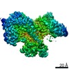

 PDBj
PDBj


 Assembly
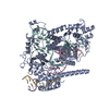


 Mass: 58.693 Da / Num. of mol.: 1 / Source method: obtained synthetically / Formula: Ni / Feature type: SUBJECT OF INVESTIGATION
 Mass: 65.409 Da / Num. of mol.: 1 / Source method: obtained synthetically / Formula: Zn / Feature type: SUBJECT OF INVESTIGATION
 Sample preparation
 Electron microscopy imaging
 FIELD EMISSION GUN / Accelerating voltage: 300 kV / Illumination mode: OTHER
 Processing