 Movie
Movie Controller

[English] 日本語
 Yorodumi
- PDB-7o9p: Crystal structure of the Awp3b (adhesin-like wall protein 3b) A-d... -
+ Open data

- Basic information
| Entry | Database: PDB / ID: 7o9p | ||||||
|---|---|---|---|---|---|---|---|
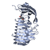
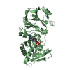
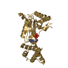
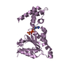
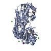
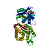
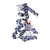
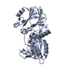
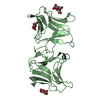
| Title | Crystal structure of the Awp3b (adhesin-like wall protein 3b) A-domain from Candida glabrata showing a gadolinium cluster | ||||||
 Components Components | AWP3b | ||||||
 Keywords Keywords | CELL ADHESION / Candida glabrata / adhesion / adhesin / Awp / adhesin-like wall protein / gadolinium / gadolinium cluster / lanthanide / lanthanide cluster / haze-protective factors / beta-helix | ||||||
| Function / homology | GADOLINIUM ATOM / AWP3b Function and homology information Function and homology information | ||||||
| Biological species |  Candida glabrata (fungus) Candida glabrata (fungus) | ||||||
| Method |  X-RAY DIFFRACTION / SYNCHROTRON / SAD / Resolution: 1.99 Å X-RAY DIFFRACTION / SYNCHROTRON / SAD / Resolution: 1.99 Å | ||||||
 Authors Authors | Reithofer, V. / de Groot, P. / Essen, L.-O. | ||||||
| Funding support |  Germany, 1items Germany, 1items
| ||||||
 Citation Citation | Journal: Plos Pathog. / Year: 2021 Title: A novel class of Candida glabrata cell wall proteins with beta-helix fold mediates adhesion in clinical isolates. Authors: Reithofer, V. / Fernandez-Pereira, J. / Alvarado, M. / de Groot, P. / Essen, L.O. | ||||||
| History |
|
- Structure visualization
| Structure viewer | Molecule: MolmilJmol/JSmol |
|---|
- Downloads & links
-Download
| PDBx/mmCIF format | 7o9p.cif.gz | 88.2 KB | Display | PDBx/mmCIF format |
|---|---|---|---|---|
| PDB format | pdb7o9p.ent.gz | 62.8 KB | Display | PDB format |
| PDBx/mmJSON format | 7o9p.json.gz | Tree view | PDBx/mmJSON format | |
| Others |  Other downloads Other downloads |
-Validation report
| Arichive directory | https://data.pdbj.org/pub/pdb/validation_reports/o9/7o9pftp://data.pdbj.org/pub/pdb/validation_reports/o9/7o9p | HTTPS FTP |
|---|
-Related structure data


-Links
 PDBj
PDBj- Assembly
| Deposited unit | 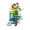
| |||||||||
|---|---|---|---|---|---|---|---|---|---|---|
| 1 |
| |||||||||
| Unit cell |
| |||||||||
| Components on special symmetry positions |
|
-Components
| #1: Protein | Mass: 38713.375 Da / Num. of mol.: 1 Source method: isolated from a genetically manipulated source Source: (gene. exp.) Candida glabrata (fungus) / Gene: AWP3b, GWK60_J11715 / Production host:  | ||||||
|---|---|---|---|---|---|---|---|
| #2: Chemical | ChemComp-GD /   Mass: 157.250 Da / Num. of mol.: 42 / Source method: obtained synthetically / Formula: Gd Mass: 157.250 Da / Num. of mol.: 42 / Source method: obtained synthetically / Formula: Gd#3: Water | ChemComp-HOH / |  Mass: 18.015 Da / Num. of mol.: 290 / Source method: isolated from a natural source / Formula: H2O Mass: 18.015 Da / Num. of mol.: 290 / Source method: isolated from a natural source / Formula: H2OHas ligand of interest | N | Has protein modification | Y | |
-Experimental details
-Experiment
| Experiment | Method: X-RAY DIFFRACTION / Number of used crystals: 1 |
|---|
- Sample preparation
| Crystal | Density Matthews: 2.95 Å3/Da / Density % sol: 58.34 % |
|---|---|
| Crystal grow | Temperature: 291.15 K / Method: vapor diffusion, sitting drop Details: 0.2 M magnesium chloride, 0.1 M Tris pH 7.0, 3.0 M sodium chloride crystals soaked with 50 mM gadolinium (III) acetate |
-Data collection
| Diffraction | Mean temperature: 100 K / Serial crystal experiment: N | ||||||||||||||||||||||||||||||
|---|---|---|---|---|---|---|---|---|---|---|---|---|---|---|---|---|---|---|---|---|---|---|---|---|---|---|---|---|---|---|---|
| Diffraction source | Source: SYNCHROTRON / Site: ESRF  / Beamline: ID29 / Wavelength: 1.71237 Å / Beamline: ID29 / Wavelength: 1.71237 Å | ||||||||||||||||||||||||||||||
| Detector | Type: DECTRIS PILATUS 6M / Detector: PIXEL / Date: Jun 25, 2017 | ||||||||||||||||||||||||||||||
| Radiation | Protocol: SINGLE WAVELENGTH / Monochromatic (M) / Laue (L): M / Scattering type: x-ray | ||||||||||||||||||||||||||||||
| Radiation wavelength | Wavelength: 1.71237 Å / Relative weight: 1 | ||||||||||||||||||||||||||||||
| Reflection | Resolution: 1.99→84.2 Å / Num. obs: 30650 / % possible obs: 99.8 % / Redundancy: 18.2 % / CC1/2: 0.998 / Rmerge(I) obs: 0.104 / Rpim(I) all: 0.025 / Rrim(I) all: 0.107 / Net I/σ(I): 18.8 | ||||||||||||||||||||||||||||||
| Reflection shell | Diffraction-ID: 1
|
- Processing
| Software |
| ||||||||||||||||||||||||||||||||||||||||||||||||||||||||||||
|---|---|---|---|---|---|---|---|---|---|---|---|---|---|---|---|---|---|---|---|---|---|---|---|---|---|---|---|---|---|---|---|---|---|---|---|---|---|---|---|---|---|---|---|---|---|---|---|---|---|---|---|---|---|---|---|---|---|---|---|---|---|
| Refinement | Method to determine structure: SAD / Resolution: 1.99→27.42 Å / Cor.coef. Fo:Fc: 0.964 / Cor.coef. Fo:Fc free: 0.948 / SU B: 3.24 / SU ML: 0.09 / Cross valid method: THROUGHOUT / σ(F): 0 / ESU R: 0.126 / ESU R Free: 0.126 / Stereochemistry target values: MAXIMUM LIKELIHOOD Details: HYDROGENS HAVE BEEN ADDED IN THE RIDING POSITIONS U VALUES : REFINED INDIVIDUALLY
| ||||||||||||||||||||||||||||||||||||||||||||||||||||||||||||
| Solvent computation | Ion probe radii: 0.8 Å / Shrinkage radii: 0.8 Å / VDW probe radii: 1.2 Å / Solvent model: MASK | ||||||||||||||||||||||||||||||||||||||||||||||||||||||||||||
| Displacement parameters | Biso max: 148.06 Å2 / Biso mean: 32.892 Å2 / Biso min: 16.47 Å2
| ||||||||||||||||||||||||||||||||||||||||||||||||||||||||||||
| Refinement step | Cycle: final / Resolution: 1.99→27.42 Å
| ||||||||||||||||||||||||||||||||||||||||||||||||||||||||||||
| Refine LS restraints |
| ||||||||||||||||||||||||||||||||||||||||||||||||||||||||||||
| LS refinement shell | Resolution: 1.99→2.04 Å / Rfactor Rfree error: 0
|
