 Movie
Movie Controller

[English] 日本語
 Yorodumi
- PDB-7o86: 1.73A X-ray crystal structure of the conserved C-terminal (CCT) o... -
+ Open data

- Basic information
| Entry | Database: PDB / ID: 7o86 | ||||||
|---|---|---|---|---|---|---|---|
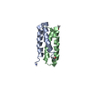
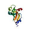
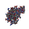
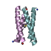
| Title | 1.73A X-ray crystal structure of the conserved C-terminal (CCT) of human SPAK | ||||||
 Components Components | STE20/SPS1-related proline-alanine-rich protein kinase | ||||||
 Keywords Keywords | SIGNALING PROTEIN / KINASE / TRANSFERASE / CCT | ||||||
| Function / homology |  Function and homology information Function and homology information: / : / positive regulation of ion transmembrane transporter activity / negative regulation of pancreatic juice secretion / chemokine (C-X-C motif) ligand 12 signaling pathway / maintenance of lens transparency / negative regulation of potassium ion transmembrane transporter activity / negative regulation of potassium ion transmembrane transport / intracellular chloride ion homeostasis / positive regulation of T cell chemotaxis ...: / : / positive regulation of ion transmembrane transporter activity / negative regulation of pancreatic juice secretion / chemokine (C-X-C motif) ligand 12 signaling pathway / maintenance of lens transparency / negative regulation of potassium ion transmembrane transporter activity / negative regulation of potassium ion transmembrane transport / intracellular chloride ion homeostasis / positive regulation of T cell chemotaxis / renal sodium ion absorption / cellular hypotonic response / macrophage activation / positive regulation of potassium ion transport / cellular response to chemokine / response to aldosterone / cellular hyperosmotic response / positive regulation of p38MAPK cascade / cell volume homeostasis / cellular response to potassium ion / response to dietary excess / peptidyl-threonine phosphorylation / sodium ion transmembrane transport / peptidyl-serine phosphorylation / regulation of blood pressure / kinase activity / protein autophosphorylation / cell body / regulation of inflammatory response / cell cortex / basolateral plasma membrane / protein phosphorylation / non-specific serine/threonine protein kinase / intracellular signal transduction / apical plasma membrane / inflammatory response / protein serine kinase activity / intracellular membrane-bounded organelle / protein serine/threonine kinase activity / protein kinase binding / signal transduction / nucleoplasm / ATP binding / cytoplasm / cytosol Similarity search - Function | ||||||
| Biological species |  Homo sapiens (human) Homo sapiens (human) | ||||||
| Method |  X-RAY DIFFRACTION / SYNCHROTRON / MOLECULAR REPLACEMENT / Resolution: 1.73 Å X-RAY DIFFRACTION / SYNCHROTRON / MOLECULAR REPLACEMENT / Resolution: 1.73 Å | ||||||
 Authors Authors | Elvers, K.T. / Bax, B.D. / Lipka-Lloyd, M. / Mehellou, Y. | ||||||
| Funding support |  United Kingdom, 1items United Kingdom, 1items
| ||||||
 Citation Citation | Journal: Chembiochem / Year: 2022 Title: Structures of the Human SPAK and OSR1 Conserved C-Terminal (CCT) Domains. Authors: Elvers, K.T. / Lipka-Lloyd, M. / Trueman, R.C. / Bax, B.D. / Mehellou, Y. | ||||||
| History |
|
- Structure visualization
| Structure viewer | Molecule: MolmilJmol/JSmol |
|---|
- Downloads & links
-Download
| PDBx/mmCIF format | 7o86.cif.gz | 73.3 KB | Display | PDBx/mmCIF format |
|---|---|---|---|---|
| PDB format | pdb7o86.ent.gz | 44.1 KB | Display | PDB format |
| PDBx/mmJSON format | 7o86.json.gz | Tree view | PDBx/mmJSON format | |
| Others |  Other downloads Other downloads |
-Validation report
| Summary document | 7o86_validation.pdf.gz | 433.6 KB | Display | wwPDB validaton report |
|---|---|---|---|---|
| Full document | 7o86_full_validation.pdf.gz | 436.4 KB | Display | |
| Data in XML | 7o86_validation.xml.gz | 12.2 KB | Display | |
| Data in CIF | 7o86_validation.cif.gz | 17.4 KB | Display | |
| Arichive directory | https://data.pdbj.org/pub/pdb/validation_reports/o8/7o86ftp://data.pdbj.org/pub/pdb/validation_reports/o8/7o86 | HTTPS FTP |
-Related structure data
| Related structure data |  7okwC  2v3sS S: Starting model for refinement C: citing same article ( |
|---|---|
| Similar structure data |


-Links
 PDBj
PDBj- Assembly
| Deposited unit | 
| ||||||||||||
|---|---|---|---|---|---|---|---|---|---|---|---|---|---|
| 1 | 
| ||||||||||||
| 2 | 
| ||||||||||||
| Unit cell |
|
-Components
| #1: Protein | Mass: 11479.870 Da / Num. of mol.: 2 Source method: isolated from a genetically manipulated source Source: (gene. exp.) Homo sapiens (human) / Gene: STK39, SPAK / Production host:  References: UniProt: Q9UEW8, non-specific serine/threonine protein kinase #2: Chemical |   Mass: 22.990 Da / Num. of mol.: 2 / Source method: obtained synthetically / Formula: Na Mass: 22.990 Da / Num. of mol.: 2 / Source method: obtained synthetically / Formula: Na#3: Chemical | ChemComp-CA / |   Mass: 40.078 Da / Num. of mol.: 1 / Source method: obtained synthetically / Formula: Ca Mass: 40.078 Da / Num. of mol.: 1 / Source method: obtained synthetically / Formula: Ca#4: Chemical | ChemComp-MG / |   Mass: 24.305 Da / Num. of mol.: 1 / Source method: obtained synthetically / Formula: Mg Mass: 24.305 Da / Num. of mol.: 1 / Source method: obtained synthetically / Formula: Mg#5: Water | ChemComp-HOH / |  Mass: 18.015 Da / Num. of mol.: 204 / Source method: isolated from a natural source / Formula: H2O Mass: 18.015 Da / Num. of mol.: 204 / Source method: isolated from a natural source / Formula: H2OHas ligand of interest | N | |
|---|
-Experimental details
-Experiment
| Experiment | Method: X-RAY DIFFRACTION / Number of used crystals: 1 |
|---|
- Sample preparation
| Crystal | Density Matthews: 2.26 Å3/Da / Density % sol: 45.65 % |
|---|---|
| Crystal grow | Temperature: 293 K / Method: vapor diffusion, sitting drop Details: 150nls Protein (1.4mgs/ml in 20 mM Tris.HCl pH 7.8, 50mM NaCl, 1mM DTT) was mixed with 50nl Morpheus A5 = 30mM Magnesium chloride hexahydrate, 30mM Calcium chloride dihydrate, 50mM Sodium ...Details: 150nls Protein (1.4mgs/ml in 20 mM Tris.HCl pH 7.8, 50mM NaCl, 1mM DTT) was mixed with 50nl Morpheus A5 = 30mM Magnesium chloride hexahydrate, 30mM Calcium chloride dihydrate, 50mM Sodium HEPES, 50mM MOPS pH 7.5, 20% v/v PEG 500* MME, 10% w/v PEG 20000. |
-Data collection
| Diffraction | Mean temperature: 100 K / Serial crystal experiment: N |
|---|---|
| Diffraction source | Source: SYNCHROTRON / Site: Diamond / Beamline: I03 / Wavelength: 0.97628 Å |
| Detector | Type: DECTRIS EIGER2 XE 16M / Detector: PIXEL / Date: Dec 15, 2020 |
| Radiation | Protocol: SINGLE WAVELENGTH / Monochromatic (M) / Laue (L): M / Scattering type: x-ray |
| Radiation wavelength | Wavelength: 0.97628 Å / Relative weight: 1 |
| Reflection | Resolution: 1.73→45.45 Å / Num. obs: 22482 / % possible obs: 99.63 % / Redundancy: 13.1 % / Biso Wilson estimate: 25.82 Å2 / CC1/2: 0.997 / CC star: 0.999 / Rpim(I) all: 0.07308 / Rrim(I) all: 0.2657 / Net I/σ(I): 6.83 |
| Reflection shell | Resolution: 1.73→1.792 Å / Redundancy: 12.8 % / Mean I/σ(I) obs: 0.43 / Num. unique obs: 2168 / CC1/2: 0.281 / CC star: 0.663 / % possible all: 97.88 |
- Processing
| Software |
| |||||||||||||||||||||||||||||||||||||||||||||||||||||||||||||||
|---|---|---|---|---|---|---|---|---|---|---|---|---|---|---|---|---|---|---|---|---|---|---|---|---|---|---|---|---|---|---|---|---|---|---|---|---|---|---|---|---|---|---|---|---|---|---|---|---|---|---|---|---|---|---|---|---|---|---|---|---|---|---|---|---|
| Refinement | Method to determine structure: MOLECULAR REPLACEMENT Starting model: 2v3s Resolution: 1.73→45.45 Å / SU ML: 0.3087 / Cross valid method: FREE R-VALUE / σ(F): 1.33 / Phase error: 28.2007 Stereochemistry target values: GeoStd + Monomer Library + CDL v1.2
| |||||||||||||||||||||||||||||||||||||||||||||||||||||||||||||||
| Solvent computation | Shrinkage radii: 0.9 Å / VDW probe radii: 1.11 Å / Solvent model: FLAT BULK SOLVENT MODEL | |||||||||||||||||||||||||||||||||||||||||||||||||||||||||||||||
| Displacement parameters | Biso mean: 34.62 Å2 | |||||||||||||||||||||||||||||||||||||||||||||||||||||||||||||||
| Refinement step | Cycle: LAST / Resolution: 1.73→45.45 Å
| |||||||||||||||||||||||||||||||||||||||||||||||||||||||||||||||
| Refine LS restraints |
| |||||||||||||||||||||||||||||||||||||||||||||||||||||||||||||||
| LS refinement shell |
|
