 Movie
Movie Controller


+ Open data

- Basic information
| Entry | Database: PDB / ID: 2q33 | |||||||||
|---|---|---|---|---|---|---|---|---|---|---|
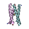
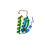
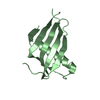
| Title | Crystal structure of all-D monellin at 1.8 A resolution | |||||||||
 Components Components |
| |||||||||
 Keywords Keywords | DE NOVO PROTEIN / ALPHA/BETA / ALL-D PROTEIN | |||||||||
| Method |  X-RAY DIFFRACTION / SYNCHROTRON / MOLECULAR REPLACEMENT / Resolution: 1.8 Å X-RAY DIFFRACTION / SYNCHROTRON / MOLECULAR REPLACEMENT / Resolution: 1.8 Å | |||||||||
 Authors Authors | Hung, L.-W. / Kohmura, M. / Ariyoshi, Y. / Kim, S.-H. | |||||||||
 Citation Citation | Journal: Acta Crystallogr.,Sect.D / Year: 1998 Title: Structure of an Enantiomeric Protein, D-Monellin at 1.8 A Resolution. Authors: Hung, L.-W. / Kohmura, M. / Ariyoshi, Y. / Kim, S.-H. | |||||||||
| History |
| |||||||||
| Remark 999 | SEQUENCE THE L-AMINO VERSION OF THE PROTEINS IN CHAINS A AND B CORRESPOND TO THE SEQUENCES IN UNP ...SEQUENCE THE L-AMINO VERSION OF THE PROTEINS IN CHAINS A AND B CORRESPOND TO THE SEQUENCES IN UNP ENTRIES MONA_DIOCU AND MONB_DIOCU RESPECTIVELY |
- Structure visualization
| Structure viewer | Molecule:  MolmilJmol/JSmol MolmilJmol/JSmol |
|---|
- Downloads & links
-Download
| PDBx/mmCIF format | 2q33.cif.gz | 35.2 KB | Display | PDBx/mmCIF format |
|---|---|---|---|---|
| PDB format | pdb2q33.ent.gz | 28 KB | Display | PDB format |
| PDBx/mmJSON format | 2q33.json.gz | Tree view | PDBx/mmJSON format | |
| Others |  Other downloads Other downloads |
-Validation report
| Arichive directory | https://data.pdbj.org/pub/pdb/validation_reports/q3/2q33ftp://data.pdbj.org/pub/pdb/validation_reports/q3/2q33 | HTTPS FTP |
|---|
-Related structure data
| Similar structure data |
|---|


-Links
 PDBj
PDBj
- Assembly
| Deposited unit | 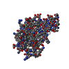
| ||||||||
|---|---|---|---|---|---|---|---|---|---|
| 1 |
| ||||||||
| Unit cell |
| ||||||||
| Components on special symmetry positions |
|
-Components
| #1: Protein/peptide | Mass: 5098.772 Da / Num. of mol.: 1 / Source method: obtained synthetically Details: The enantiomeric protein was chemically synthesized with all D-amino acids. The sequence of the protein is naturally found in Dioscoreophyllum cumminsii. |
|---|---|
| #2: Protein/peptide | Mass: 5593.396 Da / Num. of mol.: 1 / Source method: obtained synthetically Details: The enantiomeric protein was chemically synthesized with all D-amino acids. The sequence of the protein is naturally found in Dioscoreophyllum cumminsii. |
| #3: Water | ChemComp-HOH /  Mass: 18.015 Da / Num. of mol.: 82 / Source method: isolated from a natural source / Formula: H2O Mass: 18.015 Da / Num. of mol.: 82 / Source method: isolated from a natural source / Formula: H2O |
| Has protein modification | N |
-Experimental details
-Experiment
| Experiment | Method: X-RAY DIFFRACTION / Number of used crystals: 1 |
|---|
- Sample preparation
| Crystal | Density Matthews: 2.54 Å3/Da / Density % sol: 51.59 % | ||||||||||||||||||||||||
|---|---|---|---|---|---|---|---|---|---|---|---|---|---|---|---|---|---|---|---|---|---|---|---|---|---|
| Crystal grow | Temperature: 277 K / Method: vapor diffusion, sitting drop / pH: 7.2 Details: 20 MM SODIUM PHOSPHATE BUFFER, 28% PEG 8000, VAPOR DIFFUSION, SITTING DROP, TEMPERATURE 277K, pH 7.20 | ||||||||||||||||||||||||
| Crystal grow | *PLUS pH: 7.2 / Method: vapor diffusion | ||||||||||||||||||||||||
| Components of the solutions | *PLUS
|
-Data collection
| Diffraction | Mean temperature: 277 K |
|---|---|
| Diffraction source | Source: SYNCHROTRON / Site: SSRL  / Beamline: BL7-1 / Wavelength: 1.08 / Beamline: BL7-1 / Wavelength: 1.08 |
| Detector | Type: MARRESEARCH / Detector: IMAGE PLATE / Date: Sep 15, 1994 / Details: SYNCHROTRON |
| Radiation | Monochromator: SI 111 / Protocol: SINGLE WAVELENGTH / Monochromatic (M) / Laue (L): M / Scattering type: x-ray |
| Radiation wavelength | Wavelength: 1.08 Å / Relative weight: 1 |
| Reflection | Resolution: 1.8→35 Å / Num. obs: 9511 / % possible obs: 90 % / Observed criterion σ(I): 3 / Redundancy: 3.5 % / Biso Wilson estimate: 13.6 Å2 / Rmerge(I) obs: 0.042 / Net I/σ(I): 11 |
| Reflection shell | Resolution: 1.8→1.84 Å / % possible all: 70 |
| Reflection | *PLUS Highest resolution: 1.8 Å / Lowest resolution: 30 Å / Num. obs: 17490 / Rmerge(I) obs: 0.045 |
- Processing
| Software |
| ||||||||||||||||||||||||||||||||||||||||||||||||||||||||||||||||||||||||||||||||
|---|---|---|---|---|---|---|---|---|---|---|---|---|---|---|---|---|---|---|---|---|---|---|---|---|---|---|---|---|---|---|---|---|---|---|---|---|---|---|---|---|---|---|---|---|---|---|---|---|---|---|---|---|---|---|---|---|---|---|---|---|---|---|---|---|---|---|---|---|---|---|---|---|---|---|---|---|---|---|---|---|---|
| Refinement | Method to determine structure: MOLECULAR REPLACEMENT / Resolution: 1.8→6 Å / Rfactor Rfree error: 0.007 / Data cutoff high absF: 735264.36 / Data cutoff low absF: 0 / Isotropic thermal model: RESTRAINED / Cross valid method: THROUGHOUT / σ(F): 0 Details: THE PROTEIN WAS CHEMICALLY SYNTHESIZED WITH ALL D-AMINO ACIDS. THIS STRUCTURE WAS DETERMINED AND REFINED WITH L-AMINO ACID PARAMETERS. THE ACTUAL ASYMMETRIC UNIT SHOULD CONTAIN THE PROTEIN ...Details: THE PROTEIN WAS CHEMICALLY SYNTHESIZED WITH ALL D-AMINO ACIDS. THIS STRUCTURE WAS DETERMINED AND REFINED WITH L-AMINO ACID PARAMETERS. THE ACTUAL ASYMMETRIC UNIT SHOULD CONTAIN THE PROTEIN REPRESENTED BY THIS COORDINATE FILE.
| ||||||||||||||||||||||||||||||||||||||||||||||||||||||||||||||||||||||||||||||||
| Displacement parameters | Biso mean: 28.2 Å2
| ||||||||||||||||||||||||||||||||||||||||||||||||||||||||||||||||||||||||||||||||
| Refine analyze |
| ||||||||||||||||||||||||||||||||||||||||||||||||||||||||||||||||||||||||||||||||
| Refinement step | Cycle: LAST / Resolution: 1.8→6 Å
| ||||||||||||||||||||||||||||||||||||||||||||||||||||||||||||||||||||||||||||||||
| Refine LS restraints |
| ||||||||||||||||||||||||||||||||||||||||||||||||||||||||||||||||||||||||||||||||
| LS refinement shell | Resolution: 1.8→1.91 Å / Rfactor Rfree error: 0.03 / Total num. of bins used: 6
| ||||||||||||||||||||||||||||||||||||||||||||||||||||||||||||||||||||||||||||||||
| Xplor file |
| ||||||||||||||||||||||||||||||||||||||||||||||||||||||||||||||||||||||||||||||||
| Refinement | *PLUS Highest resolution: 1.8 Å / Lowest resolution: 6 Å / Num. reflection all: 18438 / Num. reflection obs: 17490 / Rfactor Rfree: 0.222 / Rfactor Rwork: 0.18 | ||||||||||||||||||||||||||||||||||||||||||||||||||||||||||||||||||||||||||||||||
| Solvent computation | *PLUS | ||||||||||||||||||||||||||||||||||||||||||||||||||||||||||||||||||||||||||||||||
| Displacement parameters | *PLUS | ||||||||||||||||||||||||||||||||||||||||||||||||||||||||||||||||||||||||||||||||
| Refine LS restraints | *PLUS
|
