 Movie
Movie Controller

[English] 日本語
 Yorodumi
- PDB-7o79: Structure of the PL6 family polysaccharide lyase Pedsa3628 from P... -
+ Open data

- Basic information
| Entry | Database: PDB / ID: 7o79 | ||||||
|---|---|---|---|---|---|---|---|
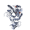
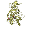
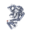
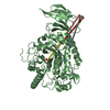
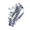
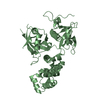
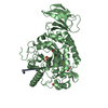
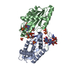
| Title | Structure of the PL6 family polysaccharide lyase Pedsa3628 from Pseudopedobacter saltans | ||||||
 Components Components | Poly(Beta-D-mannuronate) lyase | ||||||
 Keywords Keywords | LYASE / beta helix / polysaccharide lyase | ||||||
| Function / homology |  Function and homology information Function and homology informationmannuronate-specific alginate lyase / poly(beta-D-mannuronate) lyase activity Similarity search - Function | ||||||
| Biological species |  Pseudopedobacter saltans (bacteria) Pseudopedobacter saltans (bacteria) | ||||||
| Method |  X-RAY DIFFRACTION / SYNCHROTRON / MOLECULAR REPLACEMENT / Resolution: 1.93 Å X-RAY DIFFRACTION / SYNCHROTRON / MOLECULAR REPLACEMENT / Resolution: 1.93 Å | ||||||
 Authors Authors | Ballut, L. / Violot, S. / Carrique, L. / Aghajari, N. | ||||||
 Citation Citation | Journal: Glycobiology / Year: 2021 Title: Exploring molecular determinants of polysaccharide lyase family 6-1 enzyme activity. Authors: Violot, S. / Galisson, F. / Carrique, L. / Jugnarain, V. / Conchou, L. / Robert, X. / Thureau, A. / Helbert, W. / Aghajari, N. / Ballut, L. | ||||||
| History |
|
- Structure visualization
| Structure viewer | Molecule: MolmilJmol/JSmol |
|---|
- Downloads & links
-Download
| PDBx/mmCIF format | 7o79.cif.gz | 105.8 KB | Display | PDBx/mmCIF format |
|---|---|---|---|---|
| PDB format | pdb7o79.ent.gz | 76.8 KB | Display | PDB format |
| PDBx/mmJSON format | 7o79.json.gz | Tree view | PDBx/mmJSON format | |
| Others |  Other downloads Other downloads |
-Validation report
| Arichive directory | https://data.pdbj.org/pub/pdb/validation_reports/o7/7o79ftp://data.pdbj.org/pub/pdb/validation_reports/o7/7o79 | HTTPS FTP |
|---|
-Related structure data
| Related structure data | 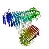 7o77C 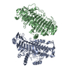 7o78C 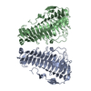 7o7aC  7o7tC 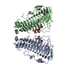 7o84C  6itgS S: Starting model for refinement C: citing same article ( |
|---|---|
| Similar structure data |


-Links
 PDBj
PDBj- Assembly
| Deposited unit | 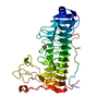
| ||||||||
|---|---|---|---|---|---|---|---|---|---|
| 1 |
| ||||||||
| Unit cell |
|
-Components
| #1: Protein | Mass: 49978.199 Da / Num. of mol.: 1 Source method: isolated from a genetically manipulated source Details: The first residue of the construct is not seen in the electron density Source: (gene. exp.) Pseudopedobacter saltans (strain ATCC 51119 / DSM 12145 / JCM 21818 / LMG 10337 / NBRC 100064 / NCIMB 13643) (bacteria)Strain: ATCC 51119 / DSM 12145 / JCM 21818 / LMG 10337 / NBRC 100064 / NCIMB 13643 Gene: Pedsa_3628 / Plasmid: pET28a / Production host: References: UniProt: F0S4Y3, mannuronate-specific alginate lyase |
|---|---|
| #2: Chemical | ChemComp-PO4 /   Mass: 94.971 Da / Num. of mol.: 1 / Source method: obtained synthetically / Formula: PO4 Mass: 94.971 Da / Num. of mol.: 1 / Source method: obtained synthetically / Formula: PO4 |
| #3: Water | ChemComp-HOH /  Mass: 18.015 Da / Num. of mol.: 344 / Source method: isolated from a natural source / Formula: H2O Mass: 18.015 Da / Num. of mol.: 344 / Source method: isolated from a natural source / Formula: H2O |
| Has ligand of interest | N |
-Experimental details
-Experiment
| Experiment | Method: X-RAY DIFFRACTION / Number of used crystals: 1 |
|---|
- Sample preparation
| Crystal | Density Matthews: 2.22 Å3/Da / Density % sol: 44.58 % |
|---|---|
| Crystal grow | Temperature: 292 K / Method: vapor diffusion, hanging drop / Details: 0.2 M K phosphate, 20% (w/v) PEG 3350 |
-Data collection
| Diffraction | Mean temperature: 100 K / Serial crystal experiment: N |
|---|---|
| Diffraction source | Source: SYNCHROTRON / Site: ESRF  / Beamline: ID30B / Wavelength: 0.95372 Å / Beamline: ID30B / Wavelength: 0.95372 Å |
| Detector | Type: DECTRIS PILATUS 6M / Detector: PIXEL / Date: Feb 19, 2018 |
| Radiation | Protocol: SINGLE WAVELENGTH / Monochromatic (M) / Laue (L): M / Scattering type: x-ray |
| Radiation wavelength | Wavelength: 0.95372 Å / Relative weight: 1 |
| Reflection | Resolution: 1.93→48.49 Å / Num. obs: 32727 / % possible obs: 98.5 % / Redundancy: 6.6 % / CC1/2: 0.996 / Net I/σ(I): 8 |
| Reflection shell | Resolution: 1.93→2.05 Å / Num. unique obs: 5026 / CC1/2: 0.54 |
- Processing
| Software |
| ||||||||||||||||||||||||||||||||||||||||||||||||||||||||||||||||||||||||||||||||||||
|---|---|---|---|---|---|---|---|---|---|---|---|---|---|---|---|---|---|---|---|---|---|---|---|---|---|---|---|---|---|---|---|---|---|---|---|---|---|---|---|---|---|---|---|---|---|---|---|---|---|---|---|---|---|---|---|---|---|---|---|---|---|---|---|---|---|---|---|---|---|---|---|---|---|---|---|---|---|---|---|---|---|---|---|---|---|
| Refinement | Method to determine structure: MOLECULAR REPLACEMENT Starting model: 6ITG Resolution: 1.93→48.49 Å / SU ML: 0.2 / Cross valid method: THROUGHOUT / σ(F): 1.34 / Phase error: 21.9 / Stereochemistry target values: ML
| ||||||||||||||||||||||||||||||||||||||||||||||||||||||||||||||||||||||||||||||||||||
| Solvent computation | Shrinkage radii: 0.9 Å / VDW probe radii: 1.11 Å / Solvent model: FLAT BULK SOLVENT MODEL | ||||||||||||||||||||||||||||||||||||||||||||||||||||||||||||||||||||||||||||||||||||
| Displacement parameters | Biso max: 64.73 Å2 / Biso mean: 29.5505 Å2 / Biso min: 15.9 Å2 | ||||||||||||||||||||||||||||||||||||||||||||||||||||||||||||||||||||||||||||||||||||
| Refinement step | Cycle: final / Resolution: 1.93→48.49 Å
| ||||||||||||||||||||||||||||||||||||||||||||||||||||||||||||||||||||||||||||||||||||
| LS refinement shell | Refine-ID: X-RAY DIFFRACTION / Rfactor Rfree error: 0 / Total num. of bins used: 11
|
