 Movie
Movie Controller

[English] 日本語
 Yorodumi
- PDB-7o7a: Structure of the PL6 family alginate lyase Pedsa0632 from Pseudop... -
+ Open data

- Basic information
| Entry | Database: PDB / ID: 7o7a | ||||||
|---|---|---|---|---|---|---|---|
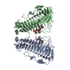
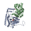
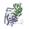
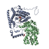
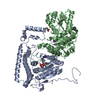
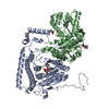
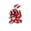
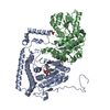
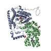
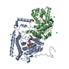
| Title | Structure of the PL6 family alginate lyase Pedsa0632 from Pseudopedobacter saltans | ||||||
 Components Components | Aliginate lyase | ||||||
 Keywords Keywords | LYASE / beta helix | ||||||
| Function / homology |  Function and homology information Function and homology informationPL-6 family / Chondroitinase B / Parallel beta-helix repeat / Parallel beta-helix repeats / Single-stranded right-handed beta-helix, Pectin lyase-like / Pectate Lyase C-like / Pectin lyase fold / Pectin lyase fold/virulence factor / 3 Solenoid / Prokaryotic membrane lipoprotein lipid attachment site profile. / Mainly Beta Similarity search - Domain/homology | ||||||
| Biological species |  Pseudopedobacter saltans (bacteria) Pseudopedobacter saltans (bacteria) | ||||||
| Method |  X-RAY DIFFRACTION / SYNCHROTRON / MOLECULAR REPLACEMENT / Resolution: 1.99 Å X-RAY DIFFRACTION / SYNCHROTRON / MOLECULAR REPLACEMENT / Resolution: 1.99 Å | ||||||
 Authors Authors | Ballut, L. / Violot, S. / Carrique, L. / Aghajari, N. | ||||||
 Citation Citation | Journal: Glycobiology / Year: 2021 Title: Exploring molecular determinants of polysaccharide lyase family 6-1 enzyme activity. Authors: Violot, S. / Galisson, F. / Carrique, L. / Jugnarain, V. / Conchou, L. / Robert, X. / Thureau, A. / Helbert, W. / Aghajari, N. / Ballut, L. | ||||||
| History |
|
- Structure visualization
| Structure viewer | Molecule: MolmilJmol/JSmol |
|---|
- Downloads & links
-Download
| PDBx/mmCIF format | 7o7a.cif.gz | 183.3 KB | Display | PDBx/mmCIF format |
|---|---|---|---|---|
| PDB format | pdb7o7a.ent.gz | 141.1 KB | Display | PDB format |
| PDBx/mmJSON format | 7o7a.json.gz | Tree view | PDBx/mmJSON format | |
| Others |  Other downloads Other downloads |
-Validation report
| Arichive directory | https://data.pdbj.org/pub/pdb/validation_reports/o7/7o7aftp://data.pdbj.org/pub/pdb/validation_reports/o7/7o7a | HTTPS FTP |
|---|
-Related structure data
| Related structure data | 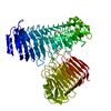 7o77C 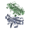 7o78C  7o79C  7o7tC  7o84C  6itgS S: Starting model for refinement C: citing same article ( |
|---|---|
| Similar structure data |
-Links
 PDBj
PDBj


- Assembly
| Deposited unit | 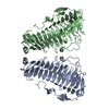
| ||||||||
|---|---|---|---|---|---|---|---|---|---|
| 1 |
| ||||||||
| Unit cell |
|
-Components
| #1: Protein | Mass: 47597.223 Da / Num. of mol.: 2 Source method: isolated from a genetically manipulated source Details: first 14 residues of construct not seen in the electron density Source: (gene. exp.) Pseudopedobacter saltans (strain ATCC 51119 / DSM 12145 / JCM 21818 / LMG 10337 / NBRC 100064 / NCIMB 13643) (bacteria)Strain: ATCC 51119 / DSM 12145 / JCM 21818 / LMG 10337 / NBRC 100064 / NCIMB 13643 Gene: Pedsa_0632 / Plasmid: pET28a / Production host: #2: Water | ChemComp-HOH / |  Mass: 18.015 Da / Num. of mol.: 610 / Source method: isolated from a natural source / Formula: H2O Mass: 18.015 Da / Num. of mol.: 610 / Source method: isolated from a natural source / Formula: H2O |
|---|
-Experimental details
-Experiment
| Experiment | Method: X-RAY DIFFRACTION / Number of used crystals: 1 |
|---|
- Sample preparation
| Crystal | Density Matthews: 2.33 Å3/Da / Density % sol: 47.3 % |
|---|---|
| Crystal grow | Temperature: 292 K / Method: vapor diffusion, hanging drop / Details: 0.2 M K sulfate, 20% (w/v) PEG 3350 |
-Data collection
| Diffraction | Mean temperature: 100 K / Serial crystal experiment: N |
|---|---|
| Diffraction source | Source: SYNCHROTRON / Site: ESRF  / Beamline: MASSIF-3 / Wavelength: 0.9677 Å / Beamline: MASSIF-3 / Wavelength: 0.9677 Å |
| Detector | Type: DECTRIS EIGER X 4M / Detector: PIXEL / Date: Dec 17, 2017 |
| Radiation | Protocol: SINGLE WAVELENGTH / Monochromatic (M) / Laue (L): M / Scattering type: x-ray |
| Radiation wavelength | Wavelength: 0.9677 Å / Relative weight: 1 |
| Reflection | Resolution: 1.99→80.49 Å / Num. obs: 58008 / % possible obs: 94.7 % / Redundancy: 6.9 % / CC1/2: 0.998 / Net I/σ(I): 11.7 |
| Reflection shell | Resolution: 1.99→2.04 Å / Num. unique obs: 4451 / CC1/2: 0.85 |
- Processing
| Software |
| ||||||||||||||||||||||||||||||||||||||||||||||||||||||||||||||||||||||||||||||||||||||||||||||||||||||||||||
|---|---|---|---|---|---|---|---|---|---|---|---|---|---|---|---|---|---|---|---|---|---|---|---|---|---|---|---|---|---|---|---|---|---|---|---|---|---|---|---|---|---|---|---|---|---|---|---|---|---|---|---|---|---|---|---|---|---|---|---|---|---|---|---|---|---|---|---|---|---|---|---|---|---|---|---|---|---|---|---|---|---|---|---|---|---|---|---|---|---|---|---|---|---|---|---|---|---|---|---|---|---|---|---|---|---|---|---|---|---|
| Refinement | Method to determine structure: MOLECULAR REPLACEMENT Starting model: 6ITG Resolution: 1.99→57.79 Å / Cor.coef. Fo:Fc: 0.924 / Cor.coef. Fo:Fc free: 0.896 / SU R Cruickshank DPI: 0.203 / Cross valid method: THROUGHOUT / σ(F): 0 / SU R Blow DPI: 0.213 / SU Rfree Blow DPI: 0.175 / SU Rfree Cruickshank DPI: 0.172
| ||||||||||||||||||||||||||||||||||||||||||||||||||||||||||||||||||||||||||||||||||||||||||||||||||||||||||||
| Displacement parameters | Biso max: 86.51 Å2 / Biso mean: 33.19 Å2 / Biso min: 16.26 Å2
| ||||||||||||||||||||||||||||||||||||||||||||||||||||||||||||||||||||||||||||||||||||||||||||||||||||||||||||
| Refine analyze | Luzzati coordinate error obs: 0.28 Å | ||||||||||||||||||||||||||||||||||||||||||||||||||||||||||||||||||||||||||||||||||||||||||||||||||||||||||||
| Refinement step | Cycle: final / Resolution: 1.99→57.79 Å
| ||||||||||||||||||||||||||||||||||||||||||||||||||||||||||||||||||||||||||||||||||||||||||||||||||||||||||||
| Refine LS restraints |
| ||||||||||||||||||||||||||||||||||||||||||||||||||||||||||||||||||||||||||||||||||||||||||||||||||||||||||||
| LS refinement shell | Resolution: 1.99→2 Å / Rfactor Rfree error: 0 / Total num. of bins used: 51
|
