 Movie
Movie Controller

[English] 日本語
 Yorodumi
- PDB-1x7y: Crystal structure of the human mitochondrial branched-chain alpha... -
+ Open data

- Basic information
| Entry | Database: PDB / ID: 1x7y | |||||||||
|---|---|---|---|---|---|---|---|---|---|---|
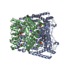
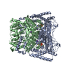
| Title | Crystal structure of the human mitochondrial branched-chain alpha-ketoacid dehydrogenase | |||||||||
 Components Components | (2-oxoisovalerate dehydrogenase ...) x 2 | |||||||||
 Keywords Keywords | OXIDOREDUCTASE / KETOACID DEHYDROGENASE / BRANCHED-CHAIN / MULTI-ENZYME COMPLEX / ACYLATION / OXIDATIVE DECARBOXYLATION MAPLE SYRUP URINE DISEASE / THIAMIN DIPHOSPHATE / PHOSPHORYLATION / FLAVOPROTEIN | |||||||||
| Function / homology |  Function and homology information Function and homology informationLoss-of-function mutations in BCKDHA or BCKDHB cause MSUD / 3-methyl-2-oxobutanoate dehydrogenase (2-methylpropanoyl-transferring) / branched-chain 2-oxo acid dehydrogenase activity / BCKDH synthesizes BCAA-CoA from KIC, KMVA, KIV / Loss-of-function mutations in DBT cause MSUD2 / Loss-of-function mutations in DLD cause MSUD3/DLDD / H139Hfs13* PPM1K causes a mild variant of MSUD / branched-chain alpha-keto acid decarboxylation to branched-chain acyl-CoA / branched-chain alpha-ketoacid dehydrogenase complex / Branched-chain ketoacid dehydrogenase kinase deficiency ...Loss-of-function mutations in BCKDHA or BCKDHB cause MSUD / 3-methyl-2-oxobutanoate dehydrogenase (2-methylpropanoyl-transferring) / branched-chain 2-oxo acid dehydrogenase activity / BCKDH synthesizes BCAA-CoA from KIC, KMVA, KIV / Loss-of-function mutations in DBT cause MSUD2 / Loss-of-function mutations in DLD cause MSUD3/DLDD / H139Hfs13* PPM1K causes a mild variant of MSUD / branched-chain alpha-keto acid decarboxylation to branched-chain acyl-CoA / branched-chain alpha-ketoacid dehydrogenase complex / Branched-chain ketoacid dehydrogenase kinase deficiency / Branched-chain amino acid catabolism / branched-chain amino acid catabolic process / carboxy-lyase activity / response to nutrient / mitochondrial matrix / nucleolus / mitochondrion / nucleoplasm / metal ion binding Similarity search - Function | |||||||||
| Biological species |  Homo sapiens (human) Homo sapiens (human) | |||||||||
| Method |  X-RAY DIFFRACTION / SYNCHROTRON / MOLECULAR REPLACEMENT / Resolution: 1.57 Å X-RAY DIFFRACTION / SYNCHROTRON / MOLECULAR REPLACEMENT / Resolution: 1.57 Å | |||||||||
 Authors Authors | Wynn, R.M. / Kato, M. / Machius, M. / Chuang, J.L. / Li, J. / Tomchick, D.R. / Chuang, D.T. | |||||||||
 Citation Citation | Journal: Structure / Year: 2004 Title: Molecular mechanism for regulation of the human mitochondrial branched-chain alpha-ketoacid dehydrogenase complex by phosphorylation Authors: Wynn, R.M. / Kato, M. / Machius, M. / Chuang, J.L. / Li, J. / Tomchick, D.R. / Chuang, D.T. #1: Journal: J.Biol.Chem. / Year: 2004Title: Crosstalk between Thiamin Diphosphate Binding and Phosphorylation Loop Conformation in Human Branched-Chain A-Ketoacid Decarboxylase/Dehydrogenase Authors: Li, J. / Wynn, R.M. / Machius, M. / Chuang, J.L. / Karthikeyan, S. / Tomchick, D.R. / Chuang, D.T. #2: Journal: J.Biol.Chem. / Year: 2003Title: Roles of His291-Alpha and His146-Beta in the Reductive Acylation Reaction Catalyzed by Human Branched-Chain Alpha-Ketoacid Dehydrogenase: Refined Phosphorylation Loop Structure in the Active Site Authors: Wynn, R.M. / Machius, M. / Chuang, J.L. / Li, J. / Tomchick, D.R. / Chuang, D.T. | |||||||||
| History |
| |||||||||
| Remark 400 | SBD MOLECULE DETAILS MOLECULE: DIHYDROLIPOYLLYSINE-RESIDUE (2-METHYLPROPANOYL) TRANSFERASE; ...SBD MOLECULE DETAILS MOLECULE: DIHYDROLIPOYLLYSINE-RESIDUE (2-METHYLPROPANOYL) TRANSFERASE; FRAGMENT: SUBUNIT-BINDING DOMAIN; EC: 2.3.1.168; GENE: BCATE2; THE SBD MOLECULE WAS CREATED FROM A GENETICALLY MODIFIED SOURCE CONSISTENT WITH THE THE SOURCE RECORDS OF THE ALPHA AND BETA SUBUNITS OF 2-OXOISOVALERATE DEHYDROGENASE FOUND IN THIS STRUCTURE. SEQUENCE: GEIKGRKTLATPAVRRLAMENNIKLSEVVGSGKDGRILKEDILNYLEKQTLEHHHHHH 1 58 RESIDUES 2-50 CORRESPOND TO RESIDUES 165-213 OF SWISSPROT ENTRY ODB2_HUMAN, ACCESSION NUMBER P11182. THE FIRST GLYCINE RESIDUE IS A CLONING ARTIFACT. THE LAST 8 C-TERMINAL RESIDUES (LEHHHHHH) ARE HIS TAG RESIDUES. | |||||||||
| Remark 999 | SEQUENCE The subunit-binding domain (SBD) of the E2 protein binds to the C-terminal region of the ...SEQUENCE The subunit-binding domain (SBD) of the E2 protein binds to the C-terminal region of the E1 beta subunit. However, the electron density of this domain is too weak to build a model, therefore this molecule has not been modeled in the coordinates. Further information on this molecule can be found in Remark 400. |
- Structure visualization
| Structure viewer | Molecule: MolmilJmol/JSmol |
|---|
- Downloads & links
-Download
| PDBx/mmCIF format | 1x7y.cif.gz | 175.8 KB | Display | PDBx/mmCIF format |
|---|---|---|---|---|
| PDB format | pdb1x7y.ent.gz | 134.2 KB | Display | PDB format |
| PDBx/mmJSON format | 1x7y.json.gz | Tree view | PDBx/mmJSON format | |
| Others |  Other downloads Other downloads |
-Validation report
| Arichive directory | https://data.pdbj.org/pub/pdb/validation_reports/x7/1x7yftp://data.pdbj.org/pub/pdb/validation_reports/x7/1x7y | HTTPS FTP |
|---|
-Related structure data
| Related structure data | 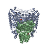 1u5bC 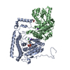 1x7wC 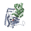 1x7xC 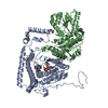 1x7zC  1x80C 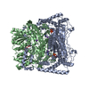 1olsS C: citing same article ( S: Starting model for refinement |
|---|---|
| Similar structure data |


-Links
 PDBj
PDBj


- Assembly
| Deposited unit | 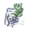
| ||||||||
|---|---|---|---|---|---|---|---|---|---|
| 1 | 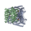
| ||||||||
| Unit cell |
| ||||||||
| Components on special symmetry positions |
| ||||||||
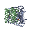
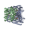
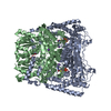
| Details | The biological assembly is a heterotetramer generated from the heterodimer in the aysmmetric unit by the operations: X-Y,-Y,2/3-Z. |
-Components
-2-oxoisovalerate dehydrogenase ... , 2 types, 2 molecules AB
| #1: Protein | Mass: 45598.121 Da / Num. of mol.: 1 / Mutation: S292N Source method: isolated from a genetically manipulated source Source: (gene. exp.) Homo sapiens (human) / Gene: BCKDHA / Plasmid: PTRCHISB / Species (production host): Escherichia coli / Production host:  References: UniProt: P12694, 3-methyl-2-oxobutanoate dehydrogenase (2-methylpropanoyl-transferring) |
|---|---|
| #2: Protein | Mass: 37902.270 Da / Num. of mol.: 1 Source method: isolated from a genetically manipulated source Source: (gene. exp.) Homo sapiens (human) / Gene: BCKDHB / Plasmid: PTRCHISB / Species (production host): Escherichia coli / Production host: References: UniProt: P21953, 3-methyl-2-oxobutanoate dehydrogenase (2-methylpropanoyl-transferring) |
-Non-polymers , 6 types, 729 molecules 


| #3: Chemical |  Mass: 39.098 Da / Num. of mol.: 2 / Source method: obtained synthetically / Formula: K Mass: 39.098 Da / Num. of mol.: 2 / Source method: obtained synthetically / Formula: K#4: Chemical | ChemComp-MN / |  Mass: 54.938 Da / Num. of mol.: 1 / Source method: obtained synthetically / Formula: Mn Mass: 54.938 Da / Num. of mol.: 1 / Source method: obtained synthetically / Formula: Mn#5: Chemical | ChemComp-CL / |  Mass: 35.453 Da / Num. of mol.: 1 / Source method: obtained synthetically / Formula: Cl Mass: 35.453 Da / Num. of mol.: 1 / Source method: obtained synthetically / Formula: Cl#6: Chemical | ChemComp-TPP / |  Mass: 425.314 Da / Num. of mol.: 1 / Source method: obtained synthetically / Formula: C12H19N4O7P2S Mass: 425.314 Da / Num. of mol.: 1 / Source method: obtained synthetically / Formula: C12H19N4O7P2S#7: Chemical | ChemComp-GOL / |  Mass: 92.094 Da / Num. of mol.: 1 / Source method: obtained synthetically / Formula: C3H8O3 Mass: 92.094 Da / Num. of mol.: 1 / Source method: obtained synthetically / Formula: C3H8O3#8: Water | ChemComp-HOH / | Mass: 18.015 Da / Num. of mol.: 723 / Source method: isolated from a natural source / Formula: H2O |
|---|
-Experimental details
-Experiment
| Experiment | Method: X-RAY DIFFRACTION / Number of used crystals: 1 |
|---|
- Sample preparation
| Crystal | Density Matthews: 2.5 Å3/Da / Density % sol: 50 % |
|---|---|
| Crystal grow | Temperature: 293 K / Method: vapor diffusion, hanging drop / pH: 5.8 Details: PEG4000, pH 5.80, VAPOR DIFFUSION, HANGING DROP, temperature 293K |
-Data collection
| Diffraction | Mean temperature: 100 K |
|---|---|
| Diffraction source | Source: SYNCHROTRON / Site: APS  / Beamline: 19-ID / Wavelength: 1.5418 / Beamline: 19-ID / Wavelength: 1.5418 |
| Detector | Type: ADSC QUANTUM 315 / Detector: CCD / Date: Oct 23, 2002 |
| Radiation | Monochromator: SI(111) / Protocol: SINGLE WAVELENGTH / Monochromatic (M) / Laue (L): M / Scattering type: x-ray |
| Radiation wavelength | Wavelength: 1.5418 Å / Relative weight: 1 |
| Reflection | Resolution: 1.57→50 Å / Num. obs: 117252 / % possible obs: 99.7 % / Observed criterion σ(I): -3 / Rmerge(I) obs: 0.051 / Net I/σ(I): 32.9 |
| Reflection shell | Resolution: 1.57→1.6 Å / Rmerge(I) obs: 0.456 / % possible all: 96.1 |
- Processing
| Software |
| ||||||||||||||||||||||||||||||||||||||||||||||||||||||||||||||||||||||||||||||||||||||||||||||||||||||||||||||||||||||||||||||||||||||||||||||||||||||||||||||||
|---|---|---|---|---|---|---|---|---|---|---|---|---|---|---|---|---|---|---|---|---|---|---|---|---|---|---|---|---|---|---|---|---|---|---|---|---|---|---|---|---|---|---|---|---|---|---|---|---|---|---|---|---|---|---|---|---|---|---|---|---|---|---|---|---|---|---|---|---|---|---|---|---|---|---|---|---|---|---|---|---|---|---|---|---|---|---|---|---|---|---|---|---|---|---|---|---|---|---|---|---|---|---|---|---|---|---|---|---|---|---|---|---|---|---|---|---|---|---|---|---|---|---|---|---|---|---|---|---|---|---|---|---|---|---|---|---|---|---|---|---|---|---|---|---|---|---|---|---|---|---|---|---|---|---|---|---|---|---|---|---|---|
| Refinement | Method to determine structure: MOLECULAR REPLACEMENT Starting model: 1OLS Resolution: 1.57→50 Å / Cor.coef. Fo:Fc: 0.975 / Cor.coef. Fo:Fc free: 0.971 / SU B: 2.462 / SU ML: 0.045 / Cross valid method: THROUGHOUT / σ(F): 0 / ESU R: 0.063 / ESU R Free: 0.062 / Stereochemistry target values: MAXIMUM LIKELIHOOD
| ||||||||||||||||||||||||||||||||||||||||||||||||||||||||||||||||||||||||||||||||||||||||||||||||||||||||||||||||||||||||||||||||||||||||||||||||||||||||||||||||
| Solvent computation | Ion probe radii: 0.8 Å / Shrinkage radii: 0.8 Å / VDW probe radii: 1.2 Å / Solvent model: MASK | ||||||||||||||||||||||||||||||||||||||||||||||||||||||||||||||||||||||||||||||||||||||||||||||||||||||||||||||||||||||||||||||||||||||||||||||||||||||||||||||||
| Displacement parameters | Biso mean: 13.511 Å2
| ||||||||||||||||||||||||||||||||||||||||||||||||||||||||||||||||||||||||||||||||||||||||||||||||||||||||||||||||||||||||||||||||||||||||||||||||||||||||||||||||
| Refinement step | Cycle: LAST / Resolution: 1.57→50 Å
| ||||||||||||||||||||||||||||||||||||||||||||||||||||||||||||||||||||||||||||||||||||||||||||||||||||||||||||||||||||||||||||||||||||||||||||||||||||||||||||||||
| Refine LS restraints |
| ||||||||||||||||||||||||||||||||||||||||||||||||||||||||||||||||||||||||||||||||||||||||||||||||||||||||||||||||||||||||||||||||||||||||||||||||||||||||||||||||
| LS refinement shell | Resolution: 1.57→1.611 Å / Total num. of bins used: 20
|
