National Institutes of Health/National Heart, Lung, and Blood Institute (NIH/NHLBI)
米国
National Institutes of Health/National Institute of Arthritis and Musculoskeletal and Skin Diseases (NIH/NIAMS)
米国
引用
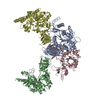
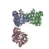
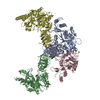
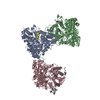
ジャーナル: Sci Adv / 年: 2021 タイトル: Structural insights into integrin αβ opening by fibronectin ligand. 著者: Stephanie Schumacher / Dirk Dedden / Roberto Vazquez Nunez / Kyoko Matoba / Junichi Takagi / Christian Biertümpfel / Naoko Mizuno / 要旨: Integrin αβ is a major fibronectin receptor critical for cell migration. Upon complex formation, fibronectin and αβ undergo conformational changes. While this is key for cell-tissue connections, ...Integrin αβ is a major fibronectin receptor critical for cell migration. Upon complex formation, fibronectin and αβ undergo conformational changes. While this is key for cell-tissue connections, its mechanism is unknown. Here, we report cryo-electron microscopy structures of native human αβ with fibronectin to 3.1-angstrom resolution, and in its resting state to 4.6-angstrom resolution. The αβ-fibronectin complex revealed simultaneous interactions at the arginine-glycine-aspartate loop, the synergy site, and a newly identified binding site proximal to adjacent to metal ion-dependent adhesion site, inducing the translocation of helix α1 to secure integrin opening. Resting αβ adopts an incompletely bent conformation, challenging the model of integrin sharp bending inhibiting ligand binding. Our biochemical and structural analyses showed that affinity of αβ for fibronectin is increased with manganese ions (Mn) while adopting the half-bent conformation, indicating that ligand-binding affinity does not depend on conformation, and αβ opening is induced by ligand-binding.
濃度: 0.15 mg/ml / 包埋: NO / シャドウイング: NO / 染色: NO / 凍結: YES 詳細: Integrin a5b1 was assembled into MSPE3D1 nanodiscs that contained bovine brain lipids (Folch fraction I) at a ratio of 1:29:3460, respectively. The assembly mix was incubated with SM-2 ...詳細: Integrin a5b1 was assembled into MSPE3D1 nanodiscs that contained bovine brain lipids (Folch fraction I) at a ratio of 1:29:3460, respectively. The assembly mix was incubated with SM-2 BioBeads to remove DDM detergent from solubilized lipids and then purified by size-exclusion chromatography using a Superose 6 3.2/300 column.
エネルギーフィルター名称: GIF Bioquantum / エネルギーフィルタースリット幅: 20 eV
-
解析
ソフトウェア
名称: PHENIX / バージョン: 1.17.1_3660: / 分類: 精密化
EMソフトウェア
ID
名称
バージョン
カテゴリ
2
SerialEM
画像取得
4
Gctf
CTF補正
7
UCSF Chimera
モデルフィッティング
8
Coot
モデルフィッティング
10
cryoSPARC
初期オイラー角割当
11
RELION
3
最終オイラー角割当
12
RELION
3
分類
13
RELION
3
3次元再構成
14
Coot
モデル精密化
15
PHENIX
1.17.1Release3660
モデル精密化
画像処理
詳細: initial image processing automatically by FOCUS pipeline: gain normalization, motion correction and dose-weighting in MotionCor2 CTF estimation by GCTF
CTF補正
タイプ: PHASE FLIPPING AND AMPLITUDE CORRECTION
粒子像の選択
選択した粒子像数: 496356 詳細: 1424 particles were manually picked and used to obtain 2D classes with distinct features as templates for Gautomatch
対称性
点対称性: C1 (非対称)
3次元再構成
解像度: 4.6 Å / 解像度の算出法: FSC 0.143 CUT-OFF / 粒子像の数: 98750 / アルゴリズム: FOURIER SPACE / クラス平均像の数: 1 / 対称性のタイプ: POINT
原子モデル構築
プロトコル: FLEXIBLE FIT / 空間: REAL
原子モデル構築
3D fitting-ID: 1 / Source name: PDB / タイプ: experimental model
 ムービー
ムービー コントローラー


 データを開く
 基本情報
基本情報 要素
要素 キーワード
キーワード 機能・相同性情報
 Homo sapiens (ヒト)
 データ登録者
 ドイツ, European Union,
  米国, 6件
  引用
引用
 構造の表示
構造の表示 ダウンロードとリンク
 その他のダウンロード


 PDBj
PDBj


 集合体
集合体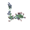

 タイプ: D-saccharide, beta linking / 分子量: 221.208 Da / 分子数: 1 / 由来タイプ: 合成 / 式: C8H15NO6

 分子量: 40.078 Da / 分子数: 7 / 由来タイプ: 組換発現 / 式: Ca
 分子量: 24.305 Da / 分子数: 1 / 由来タイプ: 合成 / 式: Mg
 試料調製
試料調製 電子顕微鏡撮影
 FIELD EMISSION GUN / 加速電圧: 300 kV / 照射モード: FLOOD BEAM
 解析
解析