National Institutes of Health/National Heart, Lung, and Blood Institute (NIH/NHLBI)
United States
National Institutes of Health/National Institute of Arthritis and Musculoskeletal and Skin Diseases (NIH/NIAMS)
United States
Citation
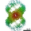
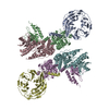
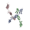
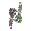
Journal: Sci Adv / Year: 2021 Title: Structural insights into integrin αβ opening by fibronectin ligand. Authors: Stephanie Schumacher / Dirk Dedden / Roberto Vazquez Nunez / Kyoko Matoba / Junichi Takagi / Christian Biertümpfel / Naoko Mizuno / Abstract: Integrin αβ is a major fibronectin receptor critical for cell migration. Upon complex formation, fibronectin and αβ undergo conformational changes. While this is key for cell-tissue connections, ...Integrin αβ is a major fibronectin receptor critical for cell migration. Upon complex formation, fibronectin and αβ undergo conformational changes. While this is key for cell-tissue connections, its mechanism is unknown. Here, we report cryo-electron microscopy structures of native human αβ with fibronectin to 3.1-angstrom resolution, and in its resting state to 4.6-angstrom resolution. The αβ-fibronectin complex revealed simultaneous interactions at the arginine-glycine-aspartate loop, the synergy site, and a newly identified binding site proximal to adjacent to metal ion-dependent adhesion site, inducing the translocation of helix α1 to secure integrin opening. Resting αβ adopts an incompletely bent conformation, challenging the model of integrin sharp bending inhibiting ligand binding. Our biochemical and structural analyses showed that affinity of αβ for fibronectin is increased with manganese ions (Mn) while adopting the half-bent conformation, indicating that ligand-binding affinity does not depend on conformation, and αβ opening is induced by ligand-binding.
History
Deposition
Mar 17, 2021
-
Header (metadata) release
Jun 2, 2021
-
Map release
Jun 2, 2021
-
Update
Nov 13, 2024
-
Current status
Nov 13, 2024
Processing site: PDBe / Status: Released
-
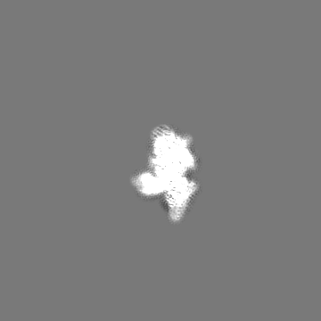
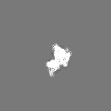
Structure visualization
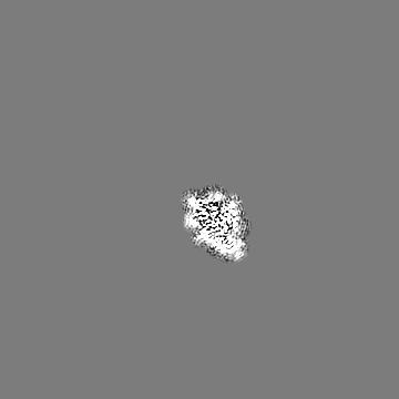
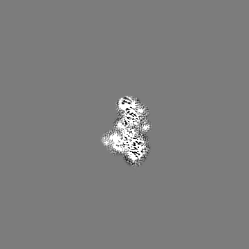
Movie
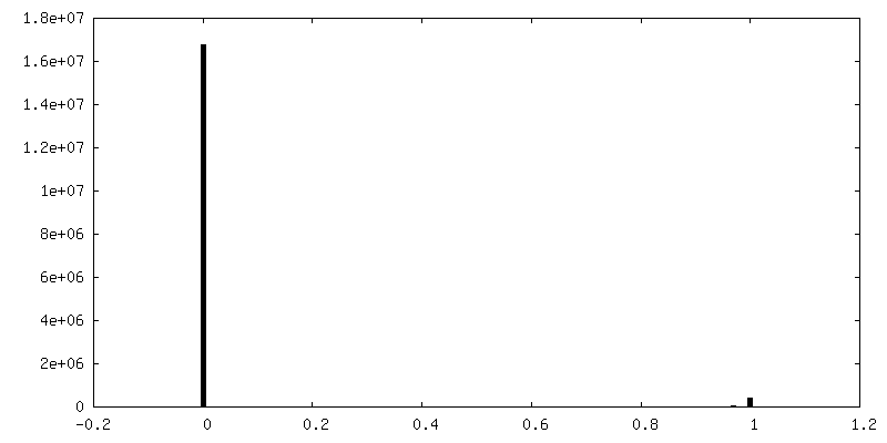
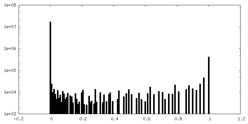
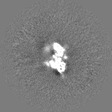
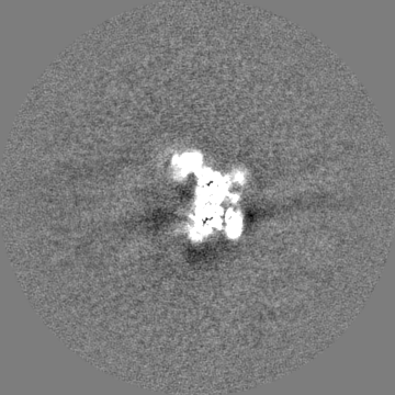
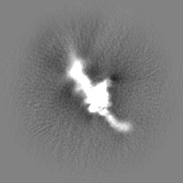
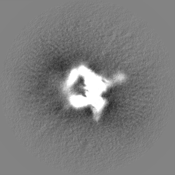
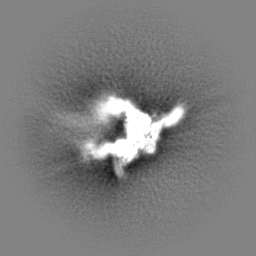
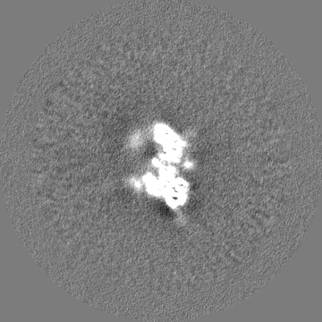
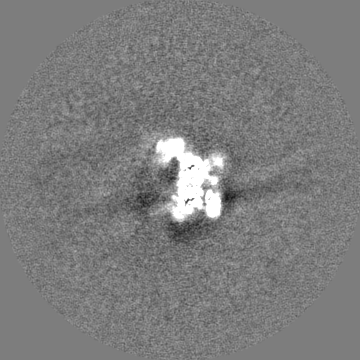
Surface view with section colored by density value
Name: MANGANESE (II) ION / type: ligand / ID: 11 / Number of copies: 7 / Formula: MN
Molecular weight
Theoretical: 54.938 Da
-
Experimental details
-
Structure determination
Method
cryo EM
Processing
single particle reconstruction
Aggregation state
particle
-
Sample preparation
Concentration
0.15 mg/mL
Buffer
pH: 7.5 Component:
Concentration
Formula
Name
50.0 mM
Tris
tris(hydroxymethyl)aminomethane
150.0 mM
NaCl
sodium chloride
1.0 mM
MnCl2
manganese chloride
Grid
Model: Quantifoil R1.2/1.3 / Material: COPPER / Mesh: 200 / Support film - Material: CARBON / Support film - topology: HOLEY / Pretreatment - Type: GLOW DISCHARGE / Pretreatment - Time: 30 sec. / Pretreatment - Atmosphere: AIR / Details: GloQube at 20 mA
Vitrification
Cryogen name: ETHANE-PROPANE / Chamber humidity: 100 % / Chamber temperature: 277 K / Instrument: FEI VITROBOT MARK IV
Details
Integrin a5b1 was assembled into MSPE3D1 nanodiscs containing lipids Folch fraction I lipids from bovine brain at a ratio of 1:29:3460, respectively. The assembly mix was incubated with SM-2 BioBeads to remove DDM detergent from solubilized lipids and then purified by size-exclusion chromatography using a Superose 6 3.2/300 column and mixed with FN7-10 and TS/2/16 at stoichiometric ratios.
-
Electron microscopy
Microscope
FEI TITAN KRIOS
Specialist optics
Energy filter - Name: GIF Bioquantum / Energy filter - Slit width: 20 eV
Image recording
Film or detector model: GATAN K3 BIOQUANTUM (6k x 4k) / Digitization - Dimensions - Width: 6144 pixel / Digitization - Dimensions - Height: 4096 pixel / Number grids imaged: 1 / Number real images: 9431 / Average exposure time: 7.39 sec. / Average electron dose: 59.7 e/Å2
Electron beam
Acceleration voltage: 300 kV / Electron source: FIELD EMISSION GUN
initial image processing automatically by FOCUS pipeline: gain normalization, motion correction and dose-weighting in MotionCor2 CTF estimation by GCTF
Particle selection
Number selected: 4218951
Startup model
Type of model: OTHER Details: 30 A-low pass filtered model from previous data analysis was used: 3754 particles were manually picked and used for 2D classification to generate a template for gautomatch, which picked ...Details: 30 A-low pass filtered model from previous data analysis was used: 3754 particles were manually picked and used for 2D classification to generate a template for gautomatch, which picked 105351 particles. Further 2D classification of 10949 classes, initial model generation and 3D in Relion yielded a prominent model from 16765 particles.
Final reconstruction
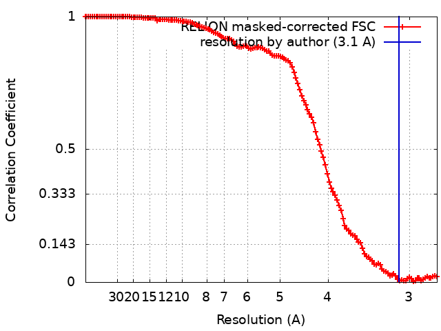
Number classes used: 2 / Applied symmetry - Point group: C1 (asymmetric) / Algorithm: FOURIER SPACE / Resolution.type: BY AUTHOR / Resolution: 3.1 Å / Resolution method: FSC 0.143 CUT-OFF / Software - Name: RELION (ver. 3.0) / Number images used: 680610
Initial angle assignment
Type: MAXIMUM LIKELIHOOD / Software - Name: RELION (ver. 3.0)
Final angle assignment
Type: MAXIMUM LIKELIHOOD / Software - Name: RELION (ver. 3.0)
Final 3D classification
Number classes: 6 / Avg.num./class: 25000 / Software - Name: RELION (ver. 3.0) / Details: 1492856 particles in total
In the structure databanks used in Yorodumi, some data are registered as the other names, "COVID-19 virus" and "2019-nCoV". Here are the details of the virus and the list of structure data.
Jan 31, 2019. EMDB accession codes are about to change! (news from PDBe EMDB page)
EMDB accession codes are about to change! (news from PDBe EMDB page)
The allocation of 4 digits for EMDB accession codes will soon come to an end. Whilst these codes will remain in use, new EMDB accession codes will include an additional digit and will expand incrementally as the available range of codes is exhausted. The current 4-digit format prefixed with “EMD-” (i.e. EMD-XXXX) will advance to a 5-digit format (i.e. EMD-XXXXX), and so on. It is currently estimated that the 4-digit codes will be depleted around Spring 2019, at which point the 5-digit format will come into force.
The EM Navigator/Yorodumi systems omit the EMD- prefix.
Related info.:Q: What is EMD? / ID/Accession-code notation in Yorodumi/EM Navigator
Yorodumi is a browser for structure data from EMDB, PDB, SASBDB, etc.
This page is also the successor to EM Navigator detail page, and also detail information page/front-end page for Omokage search.
The word "yorodu" (or yorozu) is an old Japanese word meaning "ten thousand". "mi" (miru) is to see.
Related info.:EMDB / PDB / SASBDB / Comparison of 3 databanks / Yorodumi Search / Aug 31, 2016. New EM Navigator & Yorodumi / Yorodumi Papers / Jmol/JSmol / Function and homology information / Changes in new EM Navigator and Yorodumi
 Movie
Movie Controller

 Yorodumi
 Open data
 Basic information
 Map data
 Sample
 Keywords
 Function and homology information
 Homo sapiens (human) /
 Authors
 Germany, European Union,
  United States, 6 items
  Citation
 Structure visualization
 Downloads & links
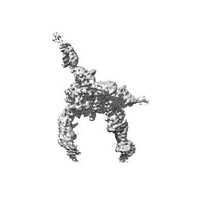 emd_12634.png
 http://ftp.pdbj.org/pub/emdb/structures/EMD-12634


 Z (Sec.)
 Y (Row.)
 X (Col.)


 Sample components
 Processing
 Electron microscopy
 FIELD EMISSION GUN