Movie
Movie Controller

[English] 日本語
 Yorodumi
- PDB-7nwl: Cryo-EM structure of human integrin alpha5beta1 (open form) in co... -
+ Open data

- Basic information
| Entry | Database: PDB / ID: 7nwl | |||||||||||||||||||||
|---|---|---|---|---|---|---|---|---|---|---|---|---|---|---|---|---|---|---|---|---|---|---|
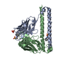
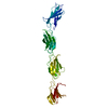
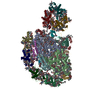
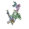
| Title | Cryo-EM structure of human integrin alpha5beta1 (open form) in complex with fibronectin and TS2/16 Fv-clasp | |||||||||||||||||||||
 Components Components |
| |||||||||||||||||||||
 Keywords Keywords | CELL ADHESION / integrin / fibronectin / TS2/16 / plasma membrane protein / a5b1 / alpha5beta1 / focal adhesion / open conformation | |||||||||||||||||||||
| Function / homology |  Function and homology information Function and homology informationintegrin alpha8-beta1 complex / myoblast fate specification / integrin alpha3-beta1 complex / integrin alpha6-beta1 complex / integrin alpha7-beta1 complex / integrin alpha10-beta1 complex / integrin alpha11-beta1 complex / positive regulation of glutamate uptake involved in transmission of nerve impulse / integrin alpha5-beta1 complex / integrin alpha9-beta1 complex ...integrin alpha8-beta1 complex / myoblast fate specification / integrin alpha3-beta1 complex / integrin alpha6-beta1 complex / integrin alpha7-beta1 complex / integrin alpha10-beta1 complex / integrin alpha11-beta1 complex / positive regulation of glutamate uptake involved in transmission of nerve impulse / integrin alpha5-beta1 complex / integrin alpha9-beta1 complex / cardiac cell fate specification / regulation of collagen catabolic process / integrin alpha1-beta1 complex / integrin binding involved in cell-matrix adhesion / cell adhesion receptor activity / integrin alpha4-beta1 complex / collagen binding involved in cell-matrix adhesion / integrin alpha2-beta1 complex / Localization of the PINCH-ILK-PARVIN complex to focal adhesions / cell-cell adhesion mediated by integrin / formation of radial glial scaffolds / negative regulation of monocyte activation / Other semaphorin interactions / Formation of the ureteric bud / negative regulation of transforming growth factor beta production / cerebellar climbing fiber to Purkinje cell synapse / positive regulation of substrate-dependent cell migration, cell attachment to substrate / CD40 signaling pathway / calcium-independent cell-matrix adhesion / positive regulation of fibroblast growth factor receptor signaling pathway / myelin sheath abaxonal region / reactive gliosis / neural crest cell migration involved in autonomic nervous system development / integrin alphav-beta1 complex / CHL1 interactions / fibrinogen complex / basement membrane organization / regulation of synapse pruning / cardiac muscle cell myoblast differentiation / RUNX2 regulates genes involved in cell migration / Fibronectin matrix formation / Attachment of bacteria to epithelial cells / alphav-beta3 integrin-vitronectin complex / MET interacts with TNS proteins / Laminin interactions / enteric nervous system development / Platelet Adhesion to exposed collagen / Developmental Lineage of Mammary Stem Cells / primordial germ cell migration / integrin activation / leukocyte tethering or rolling / ALK mutants bind TKIs / vascular endothelial growth factor receptor 2 binding / myoblast fusion / cell migration involved in sprouting angiogenesis / positive regulation of vascular endothelial growth factor signaling pathway / positive regulation of cell-substrate adhesion / Elastic fibre formation / axon extension / cardiac muscle cell differentiation / cell-substrate junction assembly / myoblast differentiation / platelet-derived growth factor receptor binding / mesodermal cell differentiation / Differentiation of Keratinocytes in Interfollicular Epidermis in Mammalian Skin / central nervous system neuron differentiation / proteoglycan binding / positive regulation of vascular endothelial growth factor receptor signaling pathway / cell projection organization / wound healing, spreading of epidermal cells / heterophilic cell-cell adhesion / positive regulation of fibroblast migration / negative regulation of Rho protein signal transduction / integrin complex / regulation of spontaneous synaptic transmission / cell adhesion mediated by integrin / MET activates PTK2 signaling / Molecules associated with elastic fibres / heterotypic cell-cell adhesion / Basigin interactions / epidermal growth factor receptor binding / lamellipodium assembly / peptidase activator activity / negative regulation of vasoconstriction / neural crest cell migration / Mechanical load activates signaling by PIEZO1 and integrins in osteocytes / extracellular matrix structural constituent / muscle organ development / Syndecan interactions / sarcomere organization / leukocyte cell-cell adhesion / p130Cas linkage to MAPK signaling for integrins / dendrite morphogenesis / positive regulation of neuroblast proliferation / biological process involved in interaction with symbiont / negative regulation of neuron differentiation / positive regulation of wound healing / response to muscle activity / positive regulation of sprouting angiogenesis / regulation of protein phosphorylation Similarity search - Function | |||||||||||||||||||||
| Biological species |  Homo sapiens (human) Homo sapiens (human) | |||||||||||||||||||||
| Method | ELECTRON MICROSCOPY / single particle reconstruction / cryo EM / Resolution: 3.1 Å | |||||||||||||||||||||
 Authors Authors | Schumacher, S. / Dedden, D. / Vazquez Nunez, R. / Matoba, K. / Takagi, J. / Biertumpfel, C. / Mizuno, N. | |||||||||||||||||||||
| Funding support |  Germany, European Union, Germany, European Union,  United States, 6items United States, 6items
| |||||||||||||||||||||
 Citation Citation | Journal: Sci Adv / Year: 2021 Title: Structural insights into integrin αβ opening by fibronectin ligand. Authors: Stephanie Schumacher / Dirk Dedden / Roberto Vazquez Nunez / Kyoko Matoba / Junichi Takagi / Christian Biertümpfel / Naoko Mizuno /  Abstract: Integrin αβ is a major fibronectin receptor critical for cell migration. Upon complex formation, fibronectin and αβ undergo conformational changes. While this is key for cell-tissue connections, ...Integrin αβ is a major fibronectin receptor critical for cell migration. Upon complex formation, fibronectin and αβ undergo conformational changes. While this is key for cell-tissue connections, its mechanism is unknown. Here, we report cryo-electron microscopy structures of native human αβ with fibronectin to 3.1-angstrom resolution, and in its resting state to 4.6-angstrom resolution. The αβ-fibronectin complex revealed simultaneous interactions at the arginine-glycine-aspartate loop, the synergy site, and a newly identified binding site proximal to adjacent to metal ion-dependent adhesion site, inducing the translocation of helix α1 to secure integrin opening. Resting αβ adopts an incompletely bent conformation, challenging the model of integrin sharp bending inhibiting ligand binding. Our biochemical and structural analyses showed that affinity of αβ for fibronectin is increased with manganese ions (Mn) while adopting the half-bent conformation, indicating that ligand-binding affinity does not depend on conformation, and αβ opening is induced by ligand-binding. | |||||||||||||||||||||
| History |
|
- Structure visualization
| Movie |
Movie viewer |
|---|---|
| Structure viewer | Molecule: MolmilJmol/JSmol |
- Downloads & links
-Download
| PDBx/mmCIF format | 7nwl.cif.gz | 343.3 KB | Display | PDBx/mmCIF format |
|---|---|---|---|---|
| PDB format | pdb7nwl.ent.gz | 262.4 KB | Display | PDB format |
| PDBx/mmJSON format | 7nwl.json.gz | Tree view | PDBx/mmJSON format | |
| Others |  Other downloads Other downloads |
-Validation report
| Arichive directory | https://data.pdbj.org/pub/pdb/validation_reports/nw/7nwlftp://data.pdbj.org/pub/pdb/validation_reports/nw/7nwl | HTTPS FTP |
|---|
-Related structure data
| Related structure data |  12634MC  7nxdC C: citing same article ( M: map data used to model this data |
|---|---|
| Similar structure data |


-Links
 PDBj
PDBj


- Assembly
| Deposited unit | 
|
|---|---|
| 1 |
|
-Components
-Protein , 3 types, 3 molecules ABC
| #1: Protein | Mass: 110111.555 Da / Num. of mol.: 1 / Source method: isolated from a natural source / Source: (natural) Homo sapiens (human) / Tissue: placenta / References: UniProt: P08648 |
|---|---|
| #2: Protein | Mass: 86338.594 Da / Num. of mol.: 1 / Source method: isolated from a natural source / Details: Sequenced used from GenBank entry CAA30790 / Source: (natural) Homo sapiens (human) / Tissue: placenta / References: UniProt: P05556 |
| #3: Protein | Mass: 39981.160 Da / Num. of mol.: 1 Source method: isolated from a genetically manipulated source Source: (gene. exp.) Homo sapiens (human) / Gene: FN1, FN / Production host:  |
-Antibody , 2 types, 2 molecules DE
| #4: Antibody | Mass: 19463.992 Da / Num. of mol.: 1 Source method: isolated from a genetically manipulated source Details: Chimera: Homo sapiens, 9606 / Source: (gene. exp.) |
|---|---|
| #5: Antibody | Mass: 18270.742 Da / Num. of mol.: 1 Source method: isolated from a genetically manipulated source Details: Chimera: Homo sapiens, 9660 / Source: (gene. exp.) |
-Sugars , 5 types, 10 molecules
| #6: Polysaccharide | beta-D-mannopyranose-(1-4)-2-acetamido-2-deoxy-beta-D-glucopyranose-(1-4)-2-acetamido-2-deoxy-beta- ...beta-D-mannopyranose-(1-4)-2-acetamido-2-deoxy-beta-D-glucopyranose-(1-4)-2-acetamido-2-deoxy-beta-D-glucopyranose Source method: isolated from a genetically manipulated source #7: Polysaccharide | Source method: isolated from a genetically manipulated source #8: Polysaccharide | alpha-D-mannopyranose-(1-3)-[alpha-D-mannopyranose-(1-6)]beta-D-mannopyranose-(1-4)-2-acetamido-2- ...alpha-D-mannopyranose-(1-3)-[alpha-D-mannopyranose-(1-6)]beta-D-mannopyranose-(1-4)-2-acetamido-2-deoxy-beta-D-glucopyranose-(1-4)-2-acetamido-2-deoxy-beta-D-glucopyranose | Source method: isolated from a genetically manipulated source #9: Polysaccharide | 2-acetamido-2-deoxy-beta-D-glucopyranose-(1-2)-alpha-D-mannopyranose-(1-6)-[alpha-D-mannopyranose- ...2-acetamido-2-deoxy-beta-D-glucopyranose-(1-2)-alpha-D-mannopyranose-(1-6)-[alpha-D-mannopyranose-(1-3)]beta-D-mannopyranose-(1-4)-2-acetamido-2-deoxy-beta-D-glucopyranose-(1-4)-2-acetamido-2-deoxy-beta-D-glucopyranose | Source method: isolated from a genetically manipulated source #10: Polysaccharide | alpha-D-mannopyranose-(1-6)-beta-D-mannopyranose-(1-4)-2-acetamido-2-deoxy-beta-D-glucopyranose-(1- ...alpha-D-mannopyranose-(1-6)-beta-D-mannopyranose-(1-4)-2-acetamido-2-deoxy-beta-D-glucopyranose-(1-4)-2-acetamido-2-deoxy-beta-D-glucopyranose | Source method: isolated from a genetically manipulated source |
|---|
-Non-polymers , 1 types, 7 molecules 
| #11: Chemical | ChemComp-MN /  Mass: 54.938 Da / Num. of mol.: 7 / Source method: obtained synthetically / Formula: Mn Mass: 54.938 Da / Num. of mol.: 7 / Source method: obtained synthetically / Formula: Mn |
|---|
-Details
| Has ligand of interest | N |
|---|---|
| Has protein modification | Y |
-Experimental details
-Experiment
| Experiment | Method: ELECTRON MICROSCOPY |
|---|---|
| EM experiment | Aggregation state: PARTICLE / 3D reconstruction method: single particle reconstruction |
- Sample preparation
| Component | Name: Ternary complex of integrin a5b1, fibronectin and TS2/16 Fv-clasp Type: COMPLEX / Entity ID: #1-#5 / Source: MULTIPLE SOURCES | ||||||||||||||||||||
|---|---|---|---|---|---|---|---|---|---|---|---|---|---|---|---|---|---|---|---|---|---|
| Molecular weight | Value: .21 MDa / Experimental value: NO | ||||||||||||||||||||
| Source (natural) | Organism: Homo sapiens (human) | ||||||||||||||||||||
| Buffer solution | pH: 7.5 | ||||||||||||||||||||
| Buffer component |
| ||||||||||||||||||||
| Specimen | Conc.: 0.15 mg/ml / Embedding applied: NO / Shadowing applied: NO / Staining applied: NO / Vitrification applied: YES Details: Integrin a5b1 was assembled into MSPE3D1 nanodiscs containing lipids Folch fraction I lipids from bovine brain at a ratio of 1:29:3460, respectively. The assembly mix was incubated with SM-2 ...Details: Integrin a5b1 was assembled into MSPE3D1 nanodiscs containing lipids Folch fraction I lipids from bovine brain at a ratio of 1:29:3460, respectively. The assembly mix was incubated with SM-2 BioBeads to remove DDM detergent from solubilized lipids and then purified by size-exclusion chromatography using a Superose 6 3.2/300 column and mixed with FN7-10 and TS/2/16 at stoichiometric ratios. | ||||||||||||||||||||
| Specimen support | Details: GloQube at 20 mA / Grid material: COPPER / Grid mesh size: 200 divisions/in. / Grid type: Quantifoil R1.2/1.3 | ||||||||||||||||||||
| Vitrification | Instrument: FEI VITROBOT MARK IV / Cryogen name: ETHANE-PROPANE / Humidity: 100 % / Chamber temperature: 277 K |
- Electron microscopy imaging
| Experimental equipment |  Model: Titan Krios / Image courtesy: FEI Company |
|---|---|
| Microscopy | Model: FEI TITAN KRIOS |
| Electron gun | Electron source:  FIELD EMISSION GUN / Accelerating voltage: 300 kV / Illumination mode: FLOOD BEAM FIELD EMISSION GUN / Accelerating voltage: 300 kV / Illumination mode: FLOOD BEAM |
| Electron lens | Mode: BRIGHT FIELD / Nominal magnification: 64000 X / Nominal defocus max: 3200 nm / Nominal defocus min: -500 nm / Cs: 2.62 mm / C2 aperture diameter: 70 µm / Alignment procedure: COMA FREE |
| Specimen holder | Cryogen: NITROGEN / Specimen holder model: FEI TITAN KRIOS AUTOGRID HOLDER |
| Image recording | Average exposure time: 7.39 sec. / Electron dose: 59.7 e/Å2 / Film or detector model: GATAN K3 BIOQUANTUM (6k x 4k) / Num. of grids imaged: 1 / Num. of real images: 9431 |
| EM imaging optics | Energyfilter name: GIF Bioquantum / Energyfilter slit width: 20 eV |
| Image scans | Sampling size: 5 µm / Width: 6144 / Height: 4096 |
- Processing
| Software | Name: PHENIX / Version: 1.17.1_3660: / Classification: refinement | ||||||||||||||||||||||||||||||||||||||||
|---|---|---|---|---|---|---|---|---|---|---|---|---|---|---|---|---|---|---|---|---|---|---|---|---|---|---|---|---|---|---|---|---|---|---|---|---|---|---|---|---|---|
| EM software |
| ||||||||||||||||||||||||||||||||||||||||
| Image processing | Details: initial image processing automatically by FOCUS pipeline: gain normalization, motion correction and dose-weighting in MotionCor2 CTF estimation by GCTF | ||||||||||||||||||||||||||||||||||||||||
| CTF correction | Type: PHASE FLIPPING AND AMPLITUDE CORRECTION | ||||||||||||||||||||||||||||||||||||||||
| Particle selection | Num. of particles selected: 4218951 | ||||||||||||||||||||||||||||||||||||||||
| Symmetry | Point symmetry: C1 (asymmetric) | ||||||||||||||||||||||||||||||||||||||||
| 3D reconstruction | Resolution: 3.1 Å / Resolution method: FSC 0.143 CUT-OFF / Num. of particles: 680610 / Algorithm: FOURIER SPACE / Num. of class averages: 2 / Symmetry type: POINT | ||||||||||||||||||||||||||||||||||||||||
| Atomic model building | Protocol: FLEXIBLE FIT / Space: REAL | ||||||||||||||||||||||||||||||||||||||||
| Atomic model building | 3D fitting-ID: 1 / Source name: PDB / Type: experimental model
| ||||||||||||||||||||||||||||||||||||||||
| Refinement | Cross valid method: NONE Stereochemistry target values: GeoStd + Monomer Library + CDL v1.2 | ||||||||||||||||||||||||||||||||||||||||
| Displacement parameters | Biso mean: 113.02 Å2 | ||||||||||||||||||||||||||||||||||||||||
| Refine LS restraints |
|