 Movie
Movie Controller

[English] 日本語
 Yorodumi
- PDB-7nwf: Crystal structure of Bacteroides thetaiotamicron EndoBT-3987 in c... -
+ Open data

- Basic information
| Entry | Database: PDB / ID: 7nwf | ||||||
|---|---|---|---|---|---|---|---|
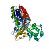
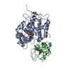
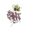
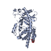
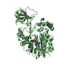
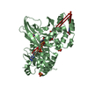
| Title | Crystal structure of Bacteroides thetaiotamicron EndoBT-3987 in complex with hybrid-type glycan (GalGlcNAcMan5GlcNAc) product | ||||||
 Components Components | Endo-beta-N-acetylglucosaminidase F1 | ||||||
 Keywords Keywords | HYDROLASE / endo-b-N-acetylglucosaminidase / EndoBT-3987 / BT3987 / glycoside hydrolase | ||||||
| Function / homology | Domain of unknown function DUF1735 / BT_3987-like, N-terminal domain / Prokaryotic membrane lipoprotein lipid attachment site profile. / Glycoside hydrolase superfamily / metal ion binding / Endo-beta-N-acetylglucosaminidase F1 Function and homology information Function and homology information | ||||||
| Biological species |  Bacteroides thetaiotaomicron (bacteria) Bacteroides thetaiotaomicron (bacteria) | ||||||
| Method |  X-RAY DIFFRACTION / SYNCHROTRON / MOLECULAR REPLACEMENT / Resolution: 2 Å X-RAY DIFFRACTION / SYNCHROTRON / MOLECULAR REPLACEMENT / Resolution: 2 Å | ||||||
 Authors Authors | Trastoy, B. / Du, J.J. / Garcia-Alija, M. / Sundberg, E.J. / Guerin, M.E. | ||||||
 Citation Citation | Journal: J.Biol.Chem. / Year: 2021 Title: GH18 endo-beta-N-acetylglucosaminidases use distinct mechanisms to process hybrid-type N-linked glycans. Authors: Trastoy, B. / Du, J.J. / Li, C. / Garcia-Alija, M. / Klontz, E.H. / Roberts, B.R. / Donahue, T.C. / Wang, L.X. / Sundberg, E.J. / Guerin, M.E. | ||||||
| History |
|
- Structure visualization
| Structure viewer | Molecule: MolmilJmol/JSmol |
|---|
- Downloads & links
-Download
| PDBx/mmCIF format | 7nwf.cif.gz | 109.7 KB | Display | PDBx/mmCIF format |
|---|---|---|---|---|
| PDB format | pdb7nwf.ent.gz | 79.8 KB | Display | PDB format |
| PDBx/mmJSON format | 7nwf.json.gz | Tree view | PDBx/mmJSON format | |
| Others |  Other downloads Other downloads |
-Validation report
| Arichive directory | https://data.pdbj.org/pub/pdb/validation_reports/nw/7nwfftp://data.pdbj.org/pub/pdb/validation_reports/nw/7nwf | HTTPS FTP |
|---|
-Related structure data
| Related structure data | 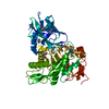 3pohS S: Starting model for refinement |
|---|---|
| Similar structure data |


-Links
 PDBj
PDBj


- Assembly
| Deposited unit | 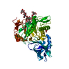
| ||||||||
|---|---|---|---|---|---|---|---|---|---|
| 1 |
| ||||||||
| Unit cell |
|
-Components
| #1: Protein | Mass: 49193.746 Da / Num. of mol.: 1 Source method: isolated from a genetically manipulated source Source: (gene. exp.) Bacteroides thetaiotaomicron (strain ATCC 29148 / DSM 2079 / NCTC 10582 / E50 / VPI-5482) (bacteria)Strain: ATCC 29148 / DSM 2079 / NCTC 10582 / E50 / VPI-5482 / Gene: BT_3987 / Production host: | ||||
|---|---|---|---|---|---|
| #2: Polysaccharide | beta-D-galactopyranose-(1-4)-2-acetamido-2-deoxy-beta-D-glucopyranose-(1-2)-alpha-D-mannopyranose- ...beta-D-galactopyranose-(1-4)-2-acetamido-2-deoxy-beta-D-glucopyranose-(1-2)-alpha-D-mannopyranose-(1-3)-[alpha-D-mannopyranose-(1-3)-[alpha-D-mannopyranose-(1-6)]alpha-D-mannopyranose-(1-6)]beta-D-mannopyranose-(1-4)-2-acetamido-2-deoxy-beta-D-glucopyranose Type: oligosaccharide / Mass: 1397.245 Da / Num. of mol.: 1 Source method: isolated from a genetically manipulated source | ||||
| #3: Chemical |   Mass: 92.094 Da / Num. of mol.: 2 / Source method: obtained synthetically / Formula: C3H8O3 Mass: 92.094 Da / Num. of mol.: 2 / Source method: obtained synthetically / Formula: C3H8O3#4: Water | ChemComp-HOH / |  Mass: 18.015 Da / Num. of mol.: 237 / Source method: isolated from a natural source / Formula: H2O Mass: 18.015 Da / Num. of mol.: 237 / Source method: isolated from a natural source / Formula: H2OHas ligand of interest | Y | |
-Experimental details
-Experiment
| Experiment | Method: X-RAY DIFFRACTION / Number of used crystals: 1 |
|---|
- Sample preparation
| Crystal | Density Matthews: 2.33 Å3/Da / Density % sol: 47.17 % |
|---|---|
| Crystal grow | Temperature: 291 K / Method: vapor diffusion, sitting drop Details: 0.2 M Sodium bromide, 0.1 M Bis-Tris propane 7.5, 20 % PEG 3350 |
-Data collection
| Diffraction | Mean temperature: 100 K / Serial crystal experiment: N |
|---|---|
| Diffraction source | Source: SYNCHROTRON / Site: Diamond  / Beamline: I24 / Wavelength: 0.9792 Å / Beamline: I24 / Wavelength: 0.9792 Å |
| Detector | Type: DECTRIS PILATUS3 6M / Detector: PIXEL / Date: Apr 6, 2019 |
| Radiation | Protocol: SINGLE WAVELENGTH / Monochromatic (M) / Laue (L): M / Scattering type: x-ray |
| Radiation wavelength | Wavelength: 0.9792 Å / Relative weight: 1 |
| Reflection | Resolution: 2→45.9 Å / Num. obs: 195095 / % possible obs: 95.06 % / Redundancy: 6.4 % / Biso Wilson estimate: 24.51 Å2 / CC1/2: 0.99 / CC star: 0.99 / Rmerge(I) obs: 0.1 / Rrim(I) all: 0.11 / Net I/σ(I): 12.58 |
| Reflection shell | Resolution: 2→2.07 Å / Redundancy: 6.4 % / Rmerge(I) obs: 0.63 / Mean I/σ(I) obs: 2.57 / Num. unique obs: 15200 / CC1/2: 0.85 / CC star: 0.96 / Rrim(I) all: 0.68 / % possible all: 76 |
- Processing
| Software |
| ||||||||||||||||||||||||||||||||||||||||||||||||||||||||||||||||||||||||
|---|---|---|---|---|---|---|---|---|---|---|---|---|---|---|---|---|---|---|---|---|---|---|---|---|---|---|---|---|---|---|---|---|---|---|---|---|---|---|---|---|---|---|---|---|---|---|---|---|---|---|---|---|---|---|---|---|---|---|---|---|---|---|---|---|---|---|---|---|---|---|---|---|---|
| Refinement | Method to determine structure: MOLECULAR REPLACEMENT Starting model: 3POH Resolution: 2→45.888 Å / SU ML: 0.22 / Cross valid method: THROUGHOUT / σ(F): 1.34 / Phase error: 21.86 / Stereochemistry target values: ML
| ||||||||||||||||||||||||||||||||||||||||||||||||||||||||||||||||||||||||
| Solvent computation | Shrinkage radii: 0.9 Å / VDW probe radii: 1.11 Å / Solvent model: FLAT BULK SOLVENT MODEL | ||||||||||||||||||||||||||||||||||||||||||||||||||||||||||||||||||||||||
| Displacement parameters | Biso max: 64.19 Å2 / Biso mean: 25.6705 Å2 / Biso min: 13.46 Å2 | ||||||||||||||||||||||||||||||||||||||||||||||||||||||||||||||||||||||||
| Refinement step | Cycle: final / Resolution: 2→45.888 Å
| ||||||||||||||||||||||||||||||||||||||||||||||||||||||||||||||||||||||||
| LS refinement shell | Refine-ID: X-RAY DIFFRACTION / Rfactor Rfree error: 0
|
