 Movie
Movie Controller

[English] 日本語
 Yorodumi
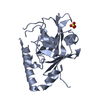
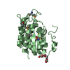
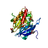
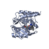
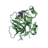
- PDB-7noy: Crystal structure of the heterocyclic toxin methyltransferase fro... -
+ Open data

- Basic information
| Entry | Database: PDB / ID: 7noy | ||||||
|---|---|---|---|---|---|---|---|
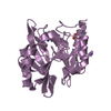
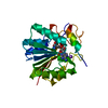
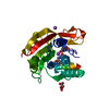
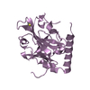
| Title | Crystal structure of the heterocyclic toxin methyltransferase from Mycobacterium tuberculosis in complex with substrate 1-hydroxyquinolin-4(1H)-one | ||||||
 Components Components | Uncharacterized protein Rv0560c | ||||||
 Keywords Keywords | TRANSFERASE / Methyltransferase / HQNO-detoxification / SAM-dependent | ||||||
| Function / homology |  Function and homology information Function and homology information2-heptyl-1-hydroxyquinolin-4(1H)-one methyltransferase / response to salicylic acid / cellular response to iron ion starvation / methyltransferase activity / methylation / response to hypoxia / cytosol Similarity search - Function | ||||||
| Biological species |   Mycobacterium tuberculosis (bacteria) Mycobacterium tuberculosis (bacteria) | ||||||
| Method |  X-RAY DIFFRACTION / SYNCHROTRON / MOLECULAR REPLACEMENT / Resolution: 1.8 Å X-RAY DIFFRACTION / SYNCHROTRON / MOLECULAR REPLACEMENT / Resolution: 1.8 Å | ||||||
 Authors Authors | Denkhaus, L. / Sartor, P. / Einsle, O. / Gerhardt, S. / Fetzner, S. | ||||||
| Funding support |  Germany, 1items Germany, 1items
| ||||||
 Citation Citation | Journal: J.Struct.Biol. / Year: 2021 Title: Structural basis of O-methylation of (2-heptyl-)1-hydroxyquinolin-4(1H)-one and related compounds by the heterocyclic toxin methyltransferase Rv0560c of Mycobacterium tuberculosis. Authors: Sartor, P. / Denkhaus, L. / Gerhardt, S. / Einsle, O. / Fetzner, S. | ||||||
| History |
|
- Structure visualization
| Structure viewer | Molecule: MolmilJmol/JSmol |
|---|
- Downloads & links
-Download
| PDBx/mmCIF format | 7noy.cif.gz | 106.5 KB | Display | PDBx/mmCIF format |
|---|---|---|---|---|
| PDB format | pdb7noy.ent.gz | 78.2 KB | Display | PDB format |
| PDBx/mmJSON format | 7noy.json.gz | Tree view | PDBx/mmJSON format | |
| Others |  Other downloads Other downloads |
-Validation report
| Arichive directory | https://data.pdbj.org/pub/pdb/validation_reports/no/7noyftp://data.pdbj.org/pub/pdb/validation_reports/no/7noy | HTTPS FTP |
|---|
-Related structure data
| Related structure data |  7bggSC  7ndmC  7nmkC S: Starting model for refinement C: citing same article ( |
|---|---|
| Similar structure data |

-Links
 PDBj
PDBj- Assembly
| Deposited unit | 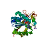
| ||||||||
|---|---|---|---|---|---|---|---|---|---|
| 1 |
| ||||||||
| Unit cell |
|
-Components
| #1: Protein | Mass: 26420.482 Da / Num. of mol.: 1 Source method: isolated from a genetically manipulated source Source: (gene. exp.) Mycobacterium tuberculosis (strain ATCC 25618 / H37Rv) (bacteria)Strain: ATCC 25618 / H37Rv / Gene: Rv0560c / Plasmid: pET28b(+) / Production host: | ||||||
|---|---|---|---|---|---|---|---|
| #2: Chemical | ChemComp-SAH /   Mass: 384.411 Da / Num. of mol.: 1 / Source method: obtained synthetically / Formula: C14H20N6O5S Mass: 384.411 Da / Num. of mol.: 1 / Source method: obtained synthetically / Formula: C14H20N6O5S | ||||||
| #3: Chemical | ChemComp-UK5 / 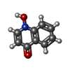  Mass: 161.157 Da / Num. of mol.: 1 / Source method: obtained synthetically / Formula: C9H7NO2 / Feature type: SUBJECT OF INVESTIGATION Mass: 161.157 Da / Num. of mol.: 1 / Source method: obtained synthetically / Formula: C9H7NO2 / Feature type: SUBJECT OF INVESTIGATION | ||||||
| #4: Chemical |   Mass: 22.990 Da / Num. of mol.: 3 / Source method: obtained synthetically / Formula: Na Mass: 22.990 Da / Num. of mol.: 3 / Source method: obtained synthetically / Formula: Na#5: Water | ChemComp-HOH / |  Mass: 18.015 Da / Num. of mol.: 282 / Source method: isolated from a natural source / Formula: H2O Mass: 18.015 Da / Num. of mol.: 282 / Source method: isolated from a natural source / Formula: H2OHas ligand of interest | Y | Has protein modification | N | |
-Experimental details
-Experiment
| Experiment | Method: X-RAY DIFFRACTION / Number of used crystals: 1 |
|---|
- Sample preparation
| Crystal | Density Matthews: 2.55 Å3/Da / Density % sol: 51.78 % |
|---|---|
| Crystal grow | Temperature: 293 K / Method: vapor diffusion, sitting drop / pH: 7 / Details: Tacsimate pH 7 |
-Data collection
| Diffraction | Mean temperature: 100 K / Serial crystal experiment: N |
|---|---|
| Diffraction source | Source: SYNCHROTRON / Site: SLS  / Beamline: X06SA / Wavelength: 1.000043 Å / Beamline: X06SA / Wavelength: 1.000043 Å |
| Detector | Type: DECTRIS EIGER X 16M / Detector: PIXEL / Date: Nov 21, 2019 |
| Radiation | Protocol: SINGLE WAVELENGTH / Monochromatic (M) / Laue (L): M / Scattering type: x-ray |
| Radiation wavelength | Wavelength: 1.000043 Å / Relative weight: 1 |
| Reflection | Resolution: 1.508→60.696 Å / Num. obs: 42031 / % possible obs: 98.1 % / Redundancy: 10.6 % / Biso Wilson estimate: 20.64 Å2 / CC1/2: 0.999 / Net I/σ(I): 19.8 |
| Reflection shell | Resolution: 1.508→1.541 Å / Num. unique obs: 2104 / CC1/2: 0.742 |
- Processing
| Software |
| ||||||||||||||||||||||||||||||||||||||||||||||||||||||||||||||||||||||||||||||||||||||||||||||||||||||||||||||||||
|---|---|---|---|---|---|---|---|---|---|---|---|---|---|---|---|---|---|---|---|---|---|---|---|---|---|---|---|---|---|---|---|---|---|---|---|---|---|---|---|---|---|---|---|---|---|---|---|---|---|---|---|---|---|---|---|---|---|---|---|---|---|---|---|---|---|---|---|---|---|---|---|---|---|---|---|---|---|---|---|---|---|---|---|---|---|---|---|---|---|---|---|---|---|---|---|---|---|---|---|---|---|---|---|---|---|---|---|---|---|---|---|---|---|---|---|
| Refinement | Method to determine structure: MOLECULAR REPLACEMENT Starting model: 7BGG Resolution: 1.8→51.16 Å / Cor.coef. Fo:Fc: 0.934 / Cor.coef. Fo:Fc free: 0.914 / SU R Cruickshank DPI: 0.111 / Cross valid method: THROUGHOUT / σ(F): 0 / SU R Blow DPI: 0.124 / SU Rfree Blow DPI: 0.117 / SU Rfree Cruickshank DPI: 0.109
| ||||||||||||||||||||||||||||||||||||||||||||||||||||||||||||||||||||||||||||||||||||||||||||||||||||||||||||||||||
| Displacement parameters | Biso mean: 20.42 Å2
| ||||||||||||||||||||||||||||||||||||||||||||||||||||||||||||||||||||||||||||||||||||||||||||||||||||||||||||||||||
| Refine analyze | Luzzati coordinate error obs: 0.19 Å | ||||||||||||||||||||||||||||||||||||||||||||||||||||||||||||||||||||||||||||||||||||||||||||||||||||||||||||||||||
| Refinement step | Cycle: 1 / Resolution: 1.8→51.16 Å
| ||||||||||||||||||||||||||||||||||||||||||||||||||||||||||||||||||||||||||||||||||||||||||||||||||||||||||||||||||
| Refine LS restraints |
| ||||||||||||||||||||||||||||||||||||||||||||||||||||||||||||||||||||||||||||||||||||||||||||||||||||||||||||||||||
| LS refinement shell | Resolution: 1.8→1.81 Å / Total num. of bins used: 50
| ||||||||||||||||||||||||||||||||||||||||||||||||||||||||||||||||||||||||||||||||||||||||||||||||||||||||||||||||||
| Refinement TLS params. | Method: refined / Origin x: 24.1921 Å / Origin y: -19.5043 Å / Origin z: -12.8439 Å
| ||||||||||||||||||||||||||||||||||||||||||||||||||||||||||||||||||||||||||||||||||||||||||||||||||||||||||||||||||
| Refinement TLS group | Selection details: { A|* } |
