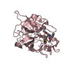
- PDB-6x0s: Structure of human plasma factor XIIa in complex with (2S)-4-(5-c... -
+
Open data
ID or keywords:
Loading...
-
Basic information
Entry
Database: PDB / ID: 6x0s
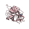
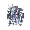
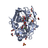
Title
Structure of human plasma factor XIIa in complex with (2S)-4-(5-chloro-1,3-benzoxazol-2-yl)-1-(N,3-dicyclohexyl-D-alanyl)-N-[(thiophen-2-yl)methyl]piperazine-2-carboxamide (compound 7)
Components
Coagulation factor XII
Keywords
BLOOD CLOTTING / preotease / inhibitor
Function / homology
Function and homology information
coagulation factor XIIa / plasma kallikrein-kinin cascade / Factor XII activation / response to misfolded protein / Defective SERPING1 causes hereditary angioedema / positive regulation of plasminogen activation / blood coagulation, intrinsic pathway / positive regulation of fibrinolysis / misfolded protein binding / zymogen activation ...coagulation factor XIIa / plasma kallikrein-kinin cascade / Factor XII activation / response to misfolded protein / Defective SERPING1 causes hereditary angioedema / positive regulation of plasminogen activation / blood coagulation, intrinsic pathway / positive regulation of fibrinolysis / misfolded protein binding / zymogen activation / Defective factor XII causes hereditary angioedema / protein autoprocessing / positive regulation of blood coagulation / fibrinolysis / : / rough endoplasmic reticulum / protein processing / blood coagulation / extracellular matrix / serine-type endopeptidase activity / innate immune response / calcium ion binding / : / extracellular exosome / extracellular region / plasma membrane Similarity search - Function
Coagulation factor XII/hepatocyte growth factor activator / Fibronectin type I domain / Fibronectin, type I / Fibronectin type-I domain signature. / Fibronectin type-I domain profile. / Fibronectin type 1 domain / Fibronectin type II domain / Fibronectin type II domain superfamily / Fibronectin type II domain / Fibronectin type-II collagen-binding domain signature. ...Coagulation factor XII/hepatocyte growth factor activator / Fibronectin type I domain / Fibronectin, type I / Fibronectin type-I domain signature. / Fibronectin type-I domain profile. / Fibronectin type 1 domain / Fibronectin type II domain / Fibronectin type II domain superfamily / Fibronectin type II domain / Fibronectin type-II collagen-binding domain signature. / Fibronectin type-II collagen-binding domain profile. / Fibronectin type 2 domain / EGF-like domain / Kringle domain / Kringle / Kringle, conserved site / Kringle superfamily / Kringle domain signature. / Kringle domain profile. / Kringle domain / EGF-like calcium-binding domain / Calcium-binding EGF-like domain / : / Kringle-like fold / Epidermal growth factor-like domain. / EGF-like domain profile. / EGF-like domain signature 1. / EGF-like domain signature 2. / EGF-like domain / Serine proteases, trypsin family, histidine active site / Serine proteases, trypsin family, serine active site / Serine proteases, trypsin family, histidine active site. / Peptidase S1A, chymotrypsin family / Serine proteases, trypsin family, serine active site. / Serine proteases, trypsin domain profile. / Trypsin-like serine protease / Serine proteases, trypsin domain / Trypsin / Trypsin-like serine proteases / Thrombin, subunit H / Peptidase S1, PA clan, chymotrypsin-like fold / Peptidase S1, PA clan / Beta Barrel / Mainly Beta Similarity search - Domain/homology
Journal: To Be Published Title: Structure of human plasma factor XIIa in complex with (2S)-4-(5-chloro-1,3-benzoxazol-2-yl)-1-(N,3-dicyclohexyl-D-alanyl)-N-[(thiophen-2-yl)methyl]piperazine-2-carboxamide (compound 7) Authors: Rao, A.U. / Chu, H.D.
Mass: 18.015 Da / Num. of mol.: 322 / Source method: isolated from a natural source / Formula: H2O
Has ligand of interest
Y
Has protein modification
Y
-
Experimental details
-
Experiment
Experiment
Method: X-RAY DIFFRACTION / Number of used crystals: 1
-
Sample preparation
Crystal
Density Matthews: 2.64 Å3/Da / Density % sol: 53.46 %
Crystal grow
Temperature: 291 K / Method: vapor diffusion, hanging drop Details: 18% PEG3350, 200 mM lithium sulfate, 0.7 mM cadmium chloride, 100 mM Bis-Tris, pH 6.3
-
Data collection
Diffraction
Mean temperature: 100 K / Serial crystal experiment: N
In the structure databanks used in Yorodumi, some data are registered as the other names, "COVID-19 virus" and "2019-nCoV". Here are the details of the virus and the list of structure data.
Jan 31, 2019. EMDB accession codes are about to change! (news from PDBe EMDB page)
EMDB accession codes are about to change! (news from PDBe EMDB page)
The allocation of 4 digits for EMDB accession codes will soon come to an end. Whilst these codes will remain in use, new EMDB accession codes will include an additional digit and will expand incrementally as the available range of codes is exhausted. The current 4-digit format prefixed with “EMD-” (i.e. EMD-XXXX) will advance to a 5-digit format (i.e. EMD-XXXXX), and so on. It is currently estimated that the 4-digit codes will be depleted around Spring 2019, at which point the 5-digit format will come into force.
The EM Navigator/Yorodumi systems omit the EMD- prefix.
Related info.:Q: What is EMD? / ID/Accession-code notation in Yorodumi/EM Navigator
Yorodumi is a browser for structure data from EMDB, PDB, SASBDB, etc.
This page is also the successor to EM Navigator detail page, and also detail information page/front-end page for Omokage search.
The word "yorodu" (or yorozu) is an old Japanese word meaning "ten thousand". "mi" (miru) is to see.
Related info.:EMDB / PDB / SASBDB / Comparison of 3 databanks / Yorodumi Search / Aug 31, 2016. New EM Navigator & Yorodumi / Yorodumi Papers / Jmol/JSmol / Function and homology information / Changes in new EM Navigator and Yorodumi
 Movie
Movie Controller

 Yorodumi
 Open data
 Basic information
 Components
 Keywords
 Function and homology information
 Homo sapiens (human)
 X-RAY DIFFRACTION /
  Authors
 Citation
 Structure visualization
 Downloads & links
 Other downloads
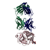


 PDBj
PDBj


 Assembly
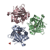

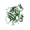

 Mass: 612.226 Da / Num. of mol.: 3 / Source method: obtained synthetically / Formula: C32H42ClN5O3S / Feature type: SUBJECT OF INVESTIGATION
 Mass: 96.063 Da / Num. of mol.: 1 / Source method: obtained synthetically / Formula: SO4
 Mass: 18.015 Da / Num. of mol.: 322 / Source method: isolated from a natural source / Formula: H2O
 Sample preparation
 / Beamline: 08ID-1 / Wavelength: 0.97949 Å
 Processing