 Movie
Movie Controller

[English] 日本語
 Yorodumi
- PDB-2wub: Crystal structure of HGFA in complex with the allosteric non- inh... -
+ Open data

- Basic information
| Entry | Database: PDB / ID: 2wub | |||||||||
|---|---|---|---|---|---|---|---|---|---|---|
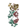
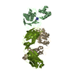
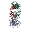
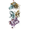
| Title | Crystal structure of HGFA in complex with the allosteric non- inhibitory antibody Fab40.deltaTrp | |||||||||
 Components Components |
| |||||||||
 Keywords Keywords | HYDROLASE/IMMUNE SYSTEM / HYDROLASE IMMUNE SYSTEM COMPLEX / DISULFIDE BOND / SERINE PROTEASE / EGF-LIKE DOMAIN / ALLOSTERIC INHIBITOR / KRINGLE / ZYMOGEN / PROTEASE / SECRETED / ANTIBODY / HYDROLASE / FAB COMPLEX / POLYMORPHISM / GLYCOPROTEIN / HYDROLASE-IMMUNE SYSTEM complex | |||||||||
| Function / homology |  Function and homology information Function and homology informationMET Receptor Activation / zymogen activation / Hydrolases; Acting on peptide bonds (peptidases); Serine endopeptidases / rough endoplasmic reticulum / serine-type peptidase activity / blood coagulation / serine-type endopeptidase activity / proteolysis / : / extracellular region / cytosol Similarity search - Function | |||||||||
| Biological species |  HOMO SAPIENS (human) HOMO SAPIENS (human) | |||||||||
| Method |  X-RAY DIFFRACTION / SYNCHROTRON / MOLECULAR REPLACEMENT / Resolution: 2.9 Å X-RAY DIFFRACTION / SYNCHROTRON / MOLECULAR REPLACEMENT / Resolution: 2.9 Å | |||||||||
 Authors Authors | Ganesan, R. / Eigenbrot, C. / Shia, S. | |||||||||
 Citation Citation | Journal: Structure / Year: 2009 Title: Unraveling the Allosteric Mechanism of Serine Protease Inhibition by an Antibody Authors: Ganesan, R. / Eigenbrot, C. / Wu, Y. / Liang, W.C. / Shia, S. / Lipari, M.T. / Kirchhofer, D. | |||||||||
| History |
| |||||||||
| Remark 700 | SHEET DETERMINATION METHOD: DSSP THE SHEETS PRESENTED AS "AA" IN EACH CHAIN ON SHEET RECORDS BELOW ... SHEET DETERMINATION METHOD: DSSP THE SHEETS PRESENTED AS "AA" IN EACH CHAIN ON SHEET RECORDS BELOW IS ACTUALLY AN 6-STRANDED BARREL THIS IS REPRESENTED BY A 7-STRANDED SHEET IN WHICH THE FIRST AND LAST STRANDS ARE IDENTICAL. THE SHEETS PRESENTED AS "AB" IN EACH CHAIN ON SHEET RECORDS BELOW IS ACTUALLY AN 6-STRANDED BARREL THIS IS REPRESENTED BY A 7-STRANDED SHEET IN WHICH THE FIRST AND LAST STRANDS ARE IDENTICAL. THE SHEETS PRESENTED AS "CA" IN EACH CHAIN ON SHEET RECORDS BELOW IS ACTUALLY AN 6-STRANDED BARREL THIS IS REPRESENTED BY A 7-STRANDED SHEET IN WHICH THE FIRST AND LAST STRANDS ARE IDENTICAL. |
- Structure visualization
| Structure viewer | Molecule: MolmilJmol/JSmol |
|---|
- Downloads & links
-Download
| PDBx/mmCIF format | 2wub.cif.gz | 267.9 KB | Display | PDBx/mmCIF format |
|---|---|---|---|---|
| PDB format | pdb2wub.ent.gz | 212.3 KB | Display | PDB format |
| PDBx/mmJSON format | 2wub.json.gz | Tree view | PDBx/mmJSON format | |
| Others |  Other downloads Other downloads |
-Validation report
| Arichive directory | https://data.pdbj.org/pub/pdb/validation_reports/wu/2wubftp://data.pdbj.org/pub/pdb/validation_reports/wu/2wub | HTTPS FTP |
|---|
-Related structure data
| Related structure data |  2wucC  3k2uC  2r0lS C: citing same article ( S: Starting model for refinement |
|---|---|
| Similar structure data |


-Links
 PDBj
PDBj


- Assembly
| Deposited unit | 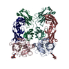
| ||||||||
|---|---|---|---|---|---|---|---|---|---|
| 1 | 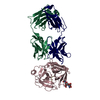
| ||||||||
| 2 | 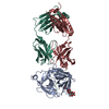
| ||||||||
| Unit cell |
|
-Components
-HEPATOCYTE GROWTH FACTOR ACTIVATOR ... , 2 types, 4 molecules ACBD
| #1: Protein | Mass: 27982.635 Da / Num. of mol.: 2 Source method: isolated from a genetically manipulated source Details: N-GLYCOSYLATION AT ASN74 / Source: (gene. exp.) HOMO SAPIENS (human) / Production host:  TRICHOPLUSIA NI (cabbage looper) TRICHOPLUSIA NI (cabbage looper)References: UniProt: Q04756, Hydrolases; Acting on peptide bonds (peptidases); Serine endopeptidases #2: Protein/peptide | Mass: 3962.654 Da / Num. of mol.: 2 Source method: isolated from a genetically manipulated source Source: (gene. exp.) HOMO SAPIENS (human) / Production host: TRICHOPLUSIA NI (cabbage looper)References: UniProt: Q04756, Hydrolases; Acting on peptide bonds (peptidases); Serine endopeptidases |
|---|
-Antibody , 2 types, 4 molecules HRLQ
| #3: Antibody | Mass: 23584.395 Da / Num. of mol.: 2 Source method: isolated from a genetically manipulated source Source: (gene. exp.) HOMO SAPIENS (human) / Production host:  #4: Antibody | Mass: 23238.748 Da / Num. of mol.: 2 Source method: isolated from a genetically manipulated source Source: (gene. exp.) HOMO SAPIENS (human) / Production host: |
|---|
-Sugars / Non-polymers , 2 types, 156 molecules 
| #5: Polysaccharide | Source method: isolated from a genetically manipulated source #6: Water | ChemComp-HOH / | Mass: 18.015 Da / Num. of mol.: 154 / Source method: isolated from a natural source / Formula: H2O |
|---|
-Details
| Has protein modification | Y |
|---|
-Experimental details
-Experiment
| Experiment | Method: X-RAY DIFFRACTION / Number of used crystals: 1 |
|---|
- Sample preparation
| Crystal | Density Matthews: 2.69 Å3/Da / Density % sol: 53.85 % / Description: NONE |
|---|---|
| Crystal grow | pH: 7.5 / Details: 10% PEG 10,000, 100 MM HEPES PH 7.5 |
-Data collection
| Diffraction | Mean temperature: 100 K |
|---|---|
| Diffraction source | Source: SYNCHROTRON / Site: ALS  / Beamline: 5.0.2 / Wavelength: 1 / Beamline: 5.0.2 / Wavelength: 1 |
| Detector | Type: ADSC CCD / Detector: CCD / Date: Mar 26, 2008 |
| Radiation | Protocol: SINGLE WAVELENGTH / Monochromatic (M) / Laue (L): M / Scattering type: x-ray |
| Radiation wavelength | Wavelength: 1 Å / Relative weight: 1 |
| Reflection | Resolution: 2.9→50 Å / Num. obs: 33747 / % possible obs: 98.8 % / Observed criterion σ(I): 2 / Redundancy: 3.7 % / Biso Wilson estimate: 113.7 Å2 / Rmerge(I) obs: 0.09 / Net I/σ(I): 14 |
| Reflection shell | Resolution: 2.9→3 Å / Redundancy: 3.6 % / Rmerge(I) obs: 0.5 / Mean I/σ(I) obs: 2.7 / % possible all: 98.3 |
- Processing
| Software |
| ||||||||||||||||||||||||||||||||||||||||||||||||||||||||||||
|---|---|---|---|---|---|---|---|---|---|---|---|---|---|---|---|---|---|---|---|---|---|---|---|---|---|---|---|---|---|---|---|---|---|---|---|---|---|---|---|---|---|---|---|---|---|---|---|---|---|---|---|---|---|---|---|---|---|---|---|---|---|
| Refinement | Method to determine structure: MOLECULAR REPLACEMENT Starting model: PDB ENTRY 2R0L Resolution: 2.9→19.99 Å / Rfactor Rfree error: 0.005 / Data cutoff high absF: 1089100.34 / Data cutoff low absF: 0 / Isotropic thermal model: RESTRAINED / Cross valid method: THROUGHOUT / σ(F): 0 / Details: BULK SOLVENT MODEL USED
| ||||||||||||||||||||||||||||||||||||||||||||||||||||||||||||
| Solvent computation | Solvent model: FLAT MODEL / Bsol: 20.5973 Å2 / ksol: 0.25 e/Å3 | ||||||||||||||||||||||||||||||||||||||||||||||||||||||||||||
| Displacement parameters | Biso mean: 70.9 Å2
| ||||||||||||||||||||||||||||||||||||||||||||||||||||||||||||
| Refine analyze |
| ||||||||||||||||||||||||||||||||||||||||||||||||||||||||||||
| Refinement step | Cycle: LAST / Resolution: 2.9→19.99 Å
| ||||||||||||||||||||||||||||||||||||||||||||||||||||||||||||
| Refine LS restraints |
| ||||||||||||||||||||||||||||||||||||||||||||||||||||||||||||
| Refine LS restraints NCS | NCS model details: CONSTR | ||||||||||||||||||||||||||||||||||||||||||||||||||||||||||||
| LS refinement shell | Resolution: 2.9→3.08 Å / Rfactor Rfree error: 0.016 / Total num. of bins used: 6
| ||||||||||||||||||||||||||||||||||||||||||||||||||||||||||||
| Xplor file |
|
