 Movie
Movie Controller

[English] 日本語
 Yorodumi
- PDB-7nms: InlB392_T332E: T332E variant of Listeria monocytogenes InlB (inte... -
+ Open data

- Basic information
| Entry | Database: PDB / ID: 7nms | ||||||
|---|---|---|---|---|---|---|---|
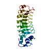
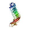
| Title | InlB392_T332E: T332E variant of Listeria monocytogenes InlB (internalin B) residues 36-392 | ||||||
 Components Components | Internalin B | ||||||
 Keywords Keywords | CELL INVASION / LEUCINE RICH REPEAT / PROTEIN BINDING / PATHOGENICITY / VIRULENCE FACTOR | ||||||
| Function / homology |  Function and homology information Function and homology informationpeptidoglycan-based cell wall / InlB-mediated entry of Listeria monocytogenes into host cell / heparin binding / lipid binding / extracellular region / plasma membrane / cytoplasm Similarity search - Function | ||||||
| Biological species |  Listeria monocytogenes serovar 1/2a (bacteria) Listeria monocytogenes serovar 1/2a (bacteria) | ||||||
| Method |  X-RAY DIFFRACTION / SYNCHROTRON / MOLECULAR REPLACEMENT / Resolution: 1.8 Å X-RAY DIFFRACTION / SYNCHROTRON / MOLECULAR REPLACEMENT / Resolution: 1.8 Å | ||||||
 Authors Authors | Geerds, C. / Niemann, H.H. | ||||||
 Citation Citation | Journal: Acta Crystallogr D Struct Biol / Year: 2022 Title: A recurring packing contact in crystals of InlB pinpoints functional binding sites in the internalin domain and the B repeat. Authors: Geerds, C. / Bleymuller, W.M. / Meyer, T. / Widmann, C. / Niemann, H.H. | ||||||
| History |
|
- Structure visualization
| Structure viewer | Molecule: MolmilJmol/JSmol |
|---|
- Downloads & links
-Download
| PDBx/mmCIF format | 7nms.cif.gz | 274.4 KB | Display | PDBx/mmCIF format |
|---|---|---|---|---|
| PDB format | pdb7nms.ent.gz | 185.6 KB | Display | PDB format |
| PDBx/mmJSON format | 7nms.json.gz | Tree view | PDBx/mmJSON format | |
| Others |  Other downloads Other downloads |
-Validation report
| Summary document | 7nms_validation.pdf.gz | 432.8 KB | Display | wwPDB validaton report |
|---|---|---|---|---|
| Full document | 7nms_full_validation.pdf.gz | 432.8 KB | Display | |
| Data in XML | 7nms_validation.xml.gz | 17.5 KB | Display | |
| Data in CIF | 7nms_validation.cif.gz | 26.4 KB | Display | |
| Arichive directory | https://data.pdbj.org/pub/pdb/validation_reports/nm/7nmsftp://data.pdbj.org/pub/pdb/validation_reports/nm/7nms | HTTPS FTP |
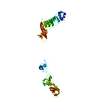
-Related structure data
| Related structure data | 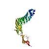 7pv8C  7pv9C 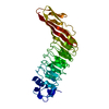 1h6tS 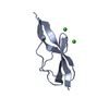 2y5pS S: Starting model for refinement C: citing same article ( |
|---|---|
| Similar structure data |


-Links
 PDBj
PDBj


- Assembly
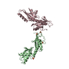
| Deposited unit | 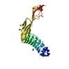
| ||||||||||||
|---|---|---|---|---|---|---|---|---|---|---|---|---|---|
| 1 |
| ||||||||||||
| Unit cell |
|
-Components
| #1: Protein | Mass: 40449.801 Da / Num. of mol.: 1 / Mutation: T332E Source method: isolated from a genetically manipulated source Details: residue 36 to 392 of InlB Source: (gene. exp.) Listeria monocytogenes serovar 1/2a (strain ATCC BAA-679 / EGD-e) (bacteria)Strain: ATCC BAA-679 / EGD-e / Gene: inlB, lmo0434 / Plasmid: PGEX-6P-1 / Production host: | ||||||||
|---|---|---|---|---|---|---|---|---|---|
| #2: Chemical |   Mass: 96.063 Da / Num. of mol.: 3 / Source method: obtained synthetically / Formula: SO4 Mass: 96.063 Da / Num. of mol.: 3 / Source method: obtained synthetically / Formula: SO4#3: Chemical |   Mass: 92.094 Da / Num. of mol.: 2 / Source method: obtained synthetically / Formula: C3H8O3 Mass: 92.094 Da / Num. of mol.: 2 / Source method: obtained synthetically / Formula: C3H8O3#4: Chemical | ChemComp-PEG / |   Mass: 106.120 Da / Num. of mol.: 1 / Source method: obtained synthetically / Formula: C4H10O3 Mass: 106.120 Da / Num. of mol.: 1 / Source method: obtained synthetically / Formula: C4H10O3#5: Water | ChemComp-HOH / |  Mass: 18.015 Da / Num. of mol.: 329 / Source method: isolated from a natural source / Formula: H2O Mass: 18.015 Da / Num. of mol.: 329 / Source method: isolated from a natural source / Formula: H2OHas ligand of interest | N | |
-Experimental details
-Experiment
| Experiment | Method: X-RAY DIFFRACTION / Number of used crystals: 1 |
|---|
- Sample preparation
| Crystal | Density Matthews: 2.43 Å3/Da / Density % sol: 49.36 % / Description: rod |
|---|---|
| Crystal grow | Temperature: 277 K / Method: vapor diffusion, sitting drop / pH: 7 Details: Reservoir solution: 0.1 M succinate, pH 7.0, 0.2 M Li2SO4, 22.5 % PEG mixture consisting of equal amounts of eight low molecular weight PEGs (PEG 300, PEG 400, PEG 500MME, PEG 550MME, PEG ...Details: Reservoir solution: 0.1 M succinate, pH 7.0, 0.2 M Li2SO4, 22.5 % PEG mixture consisting of equal amounts of eight low molecular weight PEGs (PEG 300, PEG 400, PEG 500MME, PEG 550MME, PEG 600, PEG 750MME, PEG 1,000, PEG 1,000MME); Protein solution: 10 mg/ml in 10 mM Tris, pH 8.0, 20 mM NaCl; Drop size: 100 nl + 100 nl (protein + reservoir) |
-Data collection
| Diffraction | Mean temperature: 100 K / Serial crystal experiment: N |
|---|---|
| Diffraction source | Source: SYNCHROTRON / Site: BESSY  / Beamline: 14.2 / Wavelength: 0.9184 Å / Beamline: 14.2 / Wavelength: 0.9184 Å |
| Detector | Type: DECTRIS PILATUS3 S 2M / Detector: PIXEL / Date: Jan 18, 2018 |
| Radiation | Protocol: SINGLE WAVELENGTH / Monochromatic (M) / Laue (L): M / Scattering type: x-ray |
| Radiation wavelength | Wavelength: 0.9184 Å / Relative weight: 1 |
| Reflection | Resolution: 1.8→50 Å / Num. obs: 37383 / % possible obs: 99.9 % / Redundancy: 13.2 % / Biso Wilson estimate: 30.63 Å2 / CC1/2: 1 / Rrim(I) all: 0.069 / Net I/σ(I): 23.59 |
| Reflection shell | Resolution: 1.8→1.85 Å / Redundancy: 13.5 % / Mean I/σ(I) obs: 1.88 / Num. unique obs: 2714 / CC1/2: 0.807 / Rrim(I) all: 159.9 / % possible all: 100 |
- Processing
| Software |
| |||||||||||||||||||||||||||||||||||||||||||||||||||||||||||||||||||||||||||||||||||||||||||||||||||||||||||||||||||||||||||||
|---|---|---|---|---|---|---|---|---|---|---|---|---|---|---|---|---|---|---|---|---|---|---|---|---|---|---|---|---|---|---|---|---|---|---|---|---|---|---|---|---|---|---|---|---|---|---|---|---|---|---|---|---|---|---|---|---|---|---|---|---|---|---|---|---|---|---|---|---|---|---|---|---|---|---|---|---|---|---|---|---|---|---|---|---|---|---|---|---|---|---|---|---|---|---|---|---|---|---|---|---|---|---|---|---|---|---|---|---|---|---|---|---|---|---|---|---|---|---|---|---|---|---|---|---|---|---|
| Refinement | Method to determine structure: MOLECULAR REPLACEMENT Starting model: 1h6t, 2y5p Resolution: 1.8→26.77 Å / SU ML: 0.2186 / Cross valid method: FREE R-VALUE / σ(F): 1.34 / Phase error: 24.0362 Stereochemistry target values: GeoStd + Monomer Library + CDL v1.2
| |||||||||||||||||||||||||||||||||||||||||||||||||||||||||||||||||||||||||||||||||||||||||||||||||||||||||||||||||||||||||||||
| Solvent computation | Shrinkage radii: 0.9 Å / VDW probe radii: 1.11 Å / Solvent model: FLAT BULK SOLVENT MODEL | |||||||||||||||||||||||||||||||||||||||||||||||||||||||||||||||||||||||||||||||||||||||||||||||||||||||||||||||||||||||||||||
| Displacement parameters | Biso mean: 39.6 Å2 | |||||||||||||||||||||||||||||||||||||||||||||||||||||||||||||||||||||||||||||||||||||||||||||||||||||||||||||||||||||||||||||
| Refinement step | Cycle: LAST / Resolution: 1.8→26.77 Å
| |||||||||||||||||||||||||||||||||||||||||||||||||||||||||||||||||||||||||||||||||||||||||||||||||||||||||||||||||||||||||||||
| Refine LS restraints |
| |||||||||||||||||||||||||||||||||||||||||||||||||||||||||||||||||||||||||||||||||||||||||||||||||||||||||||||||||||||||||||||
| LS refinement shell |
| |||||||||||||||||||||||||||||||||||||||||||||||||||||||||||||||||||||||||||||||||||||||||||||||||||||||||||||||||||||||||||||
| Refinement TLS params. | Method: refined / Refine-ID: X-RAY DIFFRACTION
| |||||||||||||||||||||||||||||||||||||||||||||||||||||||||||||||||||||||||||||||||||||||||||||||||||||||||||||||||||||||||||||
| Refinement TLS group | Refine-ID: X-RAY DIFFRACTION / Auth asym-ID: A / Label asym-ID: A
|
