 ムービー
ムービー コントローラー


+ データを開く

- 基本情報
基本情報
| 登録情報 | データベース: PDB / ID: 1h6t | ||||||
|---|---|---|---|---|---|---|---|
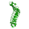
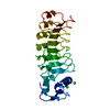
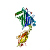
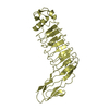
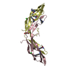
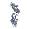
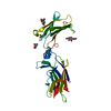
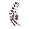
| タイトル | Internalin B: crystal structure of fused N-terminal domains. | ||||||
 要素 要素 | INTERNALIN B | ||||||
 キーワード キーワード | CELL ADHESION / LEUCINE RICH REPEAT / IG-LIKE DOMAIN / EF-HAND DOMAIN | ||||||
| 機能・相同性 |  機能・相同性情報 機能・相同性情報peptidoglycan-based cell wall / InlB-mediated entry of Listeria monocytogenes into host cell / heparin binding / lipid binding / cell surface / extracellular region / metal ion binding / plasma membrane / cytoplasm 類似検索 - 分子機能 | ||||||
| 生物種 |  LISTERIA MONOCYTOGENES (バクテリア) LISTERIA MONOCYTOGENES (バクテリア) | ||||||
| 手法 |  X線回折 / 分子置換 / 解像度: 1.6 Å X線回折 / 分子置換 / 解像度: 1.6 Å | ||||||
 データ登録者 データ登録者 | Schubert, W.-D. / Gobel, G. / Diepholz, M. / Darji, A. / Kloer, D. / Hain, T. / Chakraborty, T. / Wehland, J. / Domann, E. / Heinz, D.W. | ||||||
 引用 引用 | ジャーナル: J.Mol.Biol. / 年: 2001 タイトル: Internalins from the human pathogen Listeria monocytogenes combine three distinct folds into a contiguous internalin domain. 著者: Schubert, W.D. / Gobel, G. / Diepholz, M. / Darji, A. / Kloer, D. / Hain, T. / Chakraborty, T. / Wehland, J. / Domann, E. / Heinz, D.W. #1: ジャーナル: J.Mol.Biol. / 年: 2001タイトル: Internalins from the Human Pathogen Listeria Monocytogenes Combine Three Distinct Folds Into a Contiguous Internalin Domain 著者: Schubert, W.-D. / Gobel, G. / Diepholz, M. / Darji, A. / Kloer, D. / Hain, T. / Chakraborty, T. / Wehland, J. / Domann, E. / Heinz, D.W. | ||||||
| 履歴 |
|
- 構造の表示
構造の表示
| 構造ビューア | 分子: MolmilJmol/JSmol |
|---|
- ダウンロードとリンク
-ダウンロード
| PDBx/mmCIF形式 | 1h6t.cif.gz | 80.9 KB | 表示 | PDBx/mmCIF形式 |
|---|---|---|---|---|
| PDB形式 | pdb1h6t.ent.gz | 59.4 KB | 表示 | PDB形式 |
| PDBx/mmJSON形式 | 1h6t.json.gz | ツリー表示 | PDBx/mmJSON形式 | |
| その他 |  その他のダウンロード その他のダウンロード |
-検証レポート
| アーカイブディレクトリ | https://data.pdbj.org/pub/pdb/validation_reports/h6/1h6tftp://data.pdbj.org/pub/pdb/validation_reports/h6/1h6t | HTTPS FTP |
|---|
-関連構造データ


-リンク
 PDBj
PDBj


- 集合体
集合体
| 登録構造単位 | 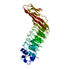
| ||||||||
|---|---|---|---|---|---|---|---|---|---|
| 1 |
| ||||||||
| 単位格子 |
|
-要素
| #1: タンパク質 | 分子量: 32395.943 Da / 分子数: 1 / 断片: LRR DOMAIN, RESIDUES 36-321 / 由来タイプ: 組換発現 由来: (組換発現) LISTERIA MONOCYTOGENES (バクテリア)株: EGD (SEROVAR 1/2A) / プラスミド: PGEX-6P-1 / 発現宿主: |
|---|---|
| #2: 水 | ChemComp-HOH /  分子量: 18.015 Da / 分子数: 475 / 由来タイプ: 天然 / 式: H2O 分子量: 18.015 Da / 分子数: 475 / 由来タイプ: 天然 / 式: H2O |
| 配列の詳細 | THE FIVE N-TERMINAL RESIDUES (GLY PRO LEU GLY SER)WERE INTRODUCED AS PART OF THE CLONING STRATEGY ...THE FIVE N-TERMINAL RESIDUES (GLY PRO LEU GLY SER)WERE INTRODUCED |
-実験情報
-実験
| 実験 | 手法: X線回折 / 使用した結晶の数: 1 |
|---|
- 試料調製
試料調製
| 結晶 | マシュー密度: 2.17 Å3/Da / 溶媒含有率: 42.8 % | |||||||||||||||||||||||||
|---|---|---|---|---|---|---|---|---|---|---|---|---|---|---|---|---|---|---|---|---|---|---|---|---|---|---|
| 結晶化 | 温度: 293 K / pH: 5 詳細: 0.2M AMMONIUM SULFATE, 0.1 M SODIUM ACETATE, PH 5.0, 13% W/V PEG 4000, 6% V/V 2-METHYL-2,4-PENTANDIOL, T=20C, PROTEIN CONC.=10 MG/ML | |||||||||||||||||||||||||
| 結晶化 | *PLUS 手法: sparse matrix method | |||||||||||||||||||||||||
| 溶液の組成 | *PLUS
|
-データ収集
| 回折 | 平均測定温度: 100 K |
|---|---|
| 放射光源 | 由来: 回転陽極 / タイプ: RIGAKU AFC-5 / 波長: 1.5418 |
| 検出器 | タイプ: RAXIS IV++ / 検出器: IMAGE PLATE / 日付: 2000年10月10日 / 詳細: OSMIC CONFOCAL MIRRORS |
| 放射 | プロトコル: SINGLE WAVELENGTH / 単色(M)・ラウエ(L): M / 散乱光タイプ: x-ray |
| 放射波長 | 波長: 1.5418 Å / 相対比: 1 |
| 反射 | 解像度: 1.6→61 Å / Num. obs: 37671 / % possible obs: 95.3 % / Observed criterion σ(I): 0 / 冗長度: 2 % / Rmerge(I) obs: 0.033 / Net I/σ(I): 22 |
| 反射 シェル | 解像度: 1.6→1.66 Å / 冗長度: 1.9 % / Rmerge(I) obs: 0.162 / Mean I/σ(I) obs: 7.42 / % possible all: 91.2 |
| 反射 | *PLUS 最低解像度: 40 Å / Num. obs: 67222 / % possible obs: 93.2 % |
| 反射 シェル | *PLUS % possible obs: 88.1 % |
- 解析
解析
| ソフトウェア |
| ||||||||||||||||||||
|---|---|---|---|---|---|---|---|---|---|---|---|---|---|---|---|---|---|---|---|---|---|
| 精密化 | 構造決定の手法: 分子置換 開始モデル: PDB ENTRY 1D0B 解像度: 1.6→62 Å / SU B: 2.22 / SU ML: 0.08 / 交差検証法: THROUGHOUT / σ(F): 0 / ESU R Free: 0.11
| ||||||||||||||||||||
| 精密化ステップ | サイクル: LAST / 解像度: 1.6→62 Å
| ||||||||||||||||||||
| ソフトウェア | *PLUS 名称: REFMAC / 分類: refinement | ||||||||||||||||||||
| 精密化 | *PLUS Rfactor obs: 0.182 / Rfactor Rfree: 0.229 | ||||||||||||||||||||
| 溶媒の処理 | *PLUS | ||||||||||||||||||||
| 原子変位パラメータ | *PLUS |
