 Movie
Movie Controller


+ Open data

- Basic information
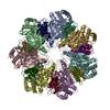
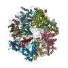
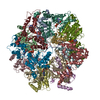
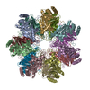
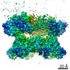
| Entry | Database: PDB / ID: 7nht | ||||||
|---|---|---|---|---|---|---|---|
| Title | Akirin2 bound human proteasome | ||||||
 Components Components |
| ||||||
 Keywords Keywords | TRANSPORT PROTEIN / proteasome / nuclear import | ||||||
| Function / homology |  Function and homology information Function and homology informationproteasome localization / regulation of muscle cell differentiation / positive regulation of B cell activation / nuclear protein quality control by the ubiquitin-proteasome system / positive regulation of adaptive immune response / embryo development ending in birth or egg hatching / purine ribonucleoside triphosphate binding / positive regulation of innate immune response / Antigen processing: Ub, ATP-independent proteasomal degradation / sperm glycocalyx ...proteasome localization / regulation of muscle cell differentiation / positive regulation of B cell activation / nuclear protein quality control by the ubiquitin-proteasome system / positive regulation of adaptive immune response / embryo development ending in birth or egg hatching / purine ribonucleoside triphosphate binding / positive regulation of innate immune response / Antigen processing: Ub, ATP-independent proteasomal degradation / sperm glycocalyx / Regulation of ornithine decarboxylase (ODC) / perinuclear theca / proteasome core complex / Proteasome assembly / Cross-presentation of soluble exogenous antigens (endosomes) / Somitogenesis / myofibril / proteasomal ubiquitin-independent protein catabolic process / sperm head-tail coupling apparatus / immune system process / proteasome endopeptidase complex / NF-kappaB binding / proteasome core complex, beta-subunit complex / threonine-type endopeptidase activity / proteasome core complex, alpha-subunit complex / proteasome assembly / transcription repressor complex / : / ciliary tip / proteasome complex / sarcomere / Regulation of activated PAK-2p34 by proteasome mediated degradation / transcription coregulator activity / Autodegradation of Cdh1 by Cdh1:APC/C / centriole / APC/C:Cdc20 mediated degradation of Securin / negative regulation of inflammatory response to antigenic stimulus / sperm end piece / Asymmetric localization of PCP proteins / Ubiquitin-dependent degradation of Cyclin D / lipopolysaccharide binding / SCF-beta-TrCP mediated degradation of Emi1 / NIK-->noncanonical NF-kB signaling / AUF1 (hnRNP D0) binds and destabilizes mRNA / TNFR2 non-canonical NF-kB pathway / P-body / Assembly of the pre-replicative complex / Vpu mediated degradation of CD4 / Cdc20:Phospho-APC/C mediated degradation of Cyclin A / Dectin-1 mediated noncanonical NF-kB signaling / Degradation of DVL / Degradation of AXIN / Degradation of CRY and PER proteins / Hh mutants are degraded by ERAD / Activation of NF-kappaB in B cells / G2/M Checkpoints / cerebral cortex development / Degradation of GLI1 by the proteasome / Hedgehog ligand biogenesis / Regulation of RUNX3 expression and activity / Autodegradation of the E3 ubiquitin ligase COP1 / Defective CFTR causes cystic fibrosis / GSK3B and BTRC:CUL1-mediated-degradation of NFE2L2 / Negative regulation of NOTCH4 signaling / APC/C:Cdh1 mediated degradation of Cdc20 and other APC/C:Cdh1 targeted proteins in late mitosis/early G1 / Hedgehog 'on' state / Vif-mediated degradation of APOBEC3G / FBXL7 down-regulates AURKA during mitotic entry and in early mitosis / positive regulation of interleukin-6 production / Degradation of GLI2 by the proteasome / GLI3 is processed to GLI3R by the proteasome / Ubiquitin-Mediated Degradation of Phosphorylated Cdc25A / MAPK6/MAPK4 signaling / Degradation of CDH1 / Degradation of beta-catenin by the destruction complex / Oxygen-dependent proline hydroxylation of Hypoxia-inducible Factor Alpha / CDK-mediated phosphorylation and removal of Cdc6 / ABC-family protein mediated transport / CLEC7A (Dectin-1) signaling / SCF(Skp2)-mediated degradation of p27/p21 / response to virus / FCERI mediated NF-kB activation / nuclear matrix / Regulation of expression of SLITs and ROBOs / Regulation of PTEN stability and activity / Interleukin-1 signaling / protein import into nucleus / Orc1 removal from chromatin / Regulation of RUNX2 expression and activity / Regulation of RAS by GAPs / The role of GTSE1 in G2/M progression after G2 checkpoint / Separation of Sister Chromatids / UCH proteinases / KEAP1-NFE2L2 pathway / peptidase activity / Downstream TCR signaling / Antigen processing: Ubiquitination & Proteasome degradation / sperm principal piece / RUNX1 regulates transcription of genes involved in differentiation of HSCs / ER-Phagosome pathway Similarity search - Function | ||||||
| Biological species |  Homo sapiens (human) Homo sapiens (human) | ||||||
| Method | ELECTRON MICROSCOPY / single particle reconstruction / cryo EM / Resolution: 3.2 Å | ||||||
 Authors Authors | Singh, K. / Brunner, H. / Grishkovskaya, I. / de Almeida, M. / Hinterndorfer, M. / Zuber, J. / Haselbach, D. | ||||||
 Citation Citation | Journal: Nature / Year: 2021 Title: AKIRIN2 controls the nuclear import of proteasomes in vertebrates. Authors: Melanie de Almeida / Matthias Hinterndorfer / Hanna Brunner / Irina Grishkovskaya / Kashish Singh / Alexander Schleiffer / Julian Jude / Sumit Deswal / Robert Kalis / Milica Vunjak / Thomas ...Authors: Melanie de Almeida / Matthias Hinterndorfer / Hanna Brunner / Irina Grishkovskaya / Kashish Singh / Alexander Schleiffer / Julian Jude / Sumit Deswal / Robert Kalis / Milica Vunjak / Thomas Lendl / Richard Imre / Elisabeth Roitinger / Tobias Neumann / Susanne Kandolf / Michael Schutzbier / Karl Mechtler / Gijs A Versteeg / David Haselbach / Johannes Zuber /     Abstract: Protein expression and turnover are controlled through a complex interplay of transcriptional, post-transcriptional and post-translational mechanisms to enable spatial and temporal regulation of ...Protein expression and turnover are controlled through a complex interplay of transcriptional, post-transcriptional and post-translational mechanisms to enable spatial and temporal regulation of cellular processes. To systematically elucidate such gene regulatory networks, we developed a CRISPR screening assay based on time-controlled Cas9 mutagenesis, intracellular immunostaining and fluorescence-activated cell sorting that enables the identification of regulatory factors independent of their effects on cellular fitness. We pioneered this approach by systematically probing the regulation of the transcription factor MYC, a master regulator of cell growth. Our screens uncover a highly conserved protein, AKIRIN2, that is essentially required for nuclear protein degradation. We found that AKIRIN2 forms homodimers that directly bind to fully assembled 20S proteasomes to mediate their nuclear import. During mitosis, proteasomes are excluded from condensing chromatin and re-imported into newly formed daughter nuclei in a highly dynamic, AKIRIN2-dependent process. Cells undergoing mitosis in the absence of AKIRIN2 become devoid of nuclear proteasomes, rapidly causing accumulation of MYC and other nuclear proteins. Collectively, our study reveals a dedicated pathway controlling the nuclear import of proteasomes in vertebrates and establishes a scalable approach to decipher regulators in essential cellular processes. | ||||||
| History |
|
- Structure visualization
| Movie |
Movie viewer |
|---|---|
| Structure viewer | Molecule: MolmilJmol/JSmol |
- Downloads & links
-Download
| PDBx/mmCIF format | 7nht.cif.gz | 570.9 KB | Display | PDBx/mmCIF format |
|---|---|---|---|---|
| PDB format | pdb7nht.ent.gz | 450.1 KB | Display | PDB format |
| PDBx/mmJSON format | 7nht.json.gz | Tree view | PDBx/mmJSON format | |
| Others |  Other downloads Other downloads |
-Validation report
| Arichive directory | https://data.pdbj.org/pub/pdb/validation_reports/nh/7nhtftp://data.pdbj.org/pub/pdb/validation_reports/nh/7nht | HTTPS FTP |
|---|
-Related structure data
| Related structure data |  12341MC C: citing same article ( M: map data used to model this data |
|---|---|
| Similar structure data | |
| EM raw data | EMPIAR-10752 (Title: Single particle cryo EM Data of a human Akirin2 proteasome complex Data size: 2.0 TB Data #1: Unaligned Multiframe micrographs of Akirin2 bound to the human proteasome [micrographs - multiframe] Data #2: Motion corrected files dose weighted and summed as well as without dose weigthening [micrographs - single frame] Data #3: Extracted particles of an Akirin2 bound proteasome [picked particles - single frame - processed]) |


-Links
 PDBj
PDBj


- Assembly
| Deposited unit | 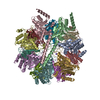
|
|---|---|
| 1 |
|
-Components
-Proteasome subunit alpha type- ... , 7 types, 7 molecules ABCDEFG
| #1: Protein | Mass: 25927.535 Da / Num. of mol.: 1 / Source method: isolated from a natural source / Source: (natural) Homo sapiens (human) / Cell line: HeLa / References: UniProt: P25787 |
|---|---|
| #2: Protein | Mass: 29525.842 Da / Num. of mol.: 1 / Source method: isolated from a natural source / Source: (natural) Homo sapiens (human) / Cell line: Hela / References: UniProt: P25789 |
| #3: Protein | Mass: 27929.891 Da / Num. of mol.: 1 / Source method: isolated from a natural source / Source: (natural) Homo sapiens (human) / Cell line: Hela / References: UniProt: O14818 |
| #4: Protein | Mass: 26435.977 Da / Num. of mol.: 1 / Source method: isolated from a natural source / Source: (natural) Homo sapiens (human) / Cell line: HeLa / References: UniProt: P28066 |
| #5: Protein | Mass: 29595.627 Da / Num. of mol.: 1 / Source method: isolated from a natural source / Source: (natural) Homo sapiens (human) / Cell line: HeLa / References: UniProt: P25786 |
| #6: Protein | Mass: 28469.252 Da / Num. of mol.: 1 / Source method: isolated from a natural source / Source: (natural) Homo sapiens (human) / Cell line: HeLa / References: UniProt: P25788 |
| #7: Protein | Mass: 27432.459 Da / Num. of mol.: 1 / Source method: isolated from a natural source / Source: (natural) Homo sapiens (human) / Cell line: HeLa / References: UniProt: P60900 |
-Proteasome subunit beta type- ... , 7 types, 7 molecules HIJKLMN
| #8: Protein | Mass: 30000.418 Da / Num. of mol.: 1 / Source method: isolated from a natural source / Source: (natural) Homo sapiens (human) / Cell line: HeLaReferences: UniProt: Q99436, proteasome endopeptidase complex |
|---|---|
| #9: Protein | Mass: 22972.896 Da / Num. of mol.: 1 / Source method: isolated from a natural source / Source: (natural) Homo sapiens (human) / Cell line: HeLaReferences: UniProt: P49720, proteasome endopeptidase complex |
| #10: Protein | Mass: 22864.277 Da / Num. of mol.: 1 / Source method: isolated from a natural source / Source: (natural) Homo sapiens (human) / Cell line: HeLaReferences: UniProt: P49721, proteasome endopeptidase complex |
| #11: Protein | Mass: 28510.248 Da / Num. of mol.: 1 / Source method: isolated from a natural source / Source: (natural) Homo sapiens (human) / Cell line: HeLaReferences: UniProt: P28074, proteasome endopeptidase complex |
| #12: Protein | Mass: 26522.396 Da / Num. of mol.: 1 / Source method: isolated from a natural source / Source: (natural) Homo sapiens (human) / Cell line: HeLaReferences: UniProt: P20618, proteasome endopeptidase complex |
| #13: Protein | Mass: 29231.178 Da / Num. of mol.: 1 / Source method: isolated from a natural source / Source: (natural) Homo sapiens (human) / Cell line: HeLaReferences: UniProt: P28070, proteasome endopeptidase complex |
| #14: Protein | Mass: 25377.652 Da / Num. of mol.: 1 / Source method: isolated from a natural source / Source: (natural) Homo sapiens (human) / Cell line: HeLaReferences: UniProt: P28072, proteasome endopeptidase complex |
-Protein / Non-polymers , 2 types, 5 molecules cd
| #15: Protein | Mass: 22525.582 Da / Num. of mol.: 2 Source method: isolated from a genetically manipulated source Source: (gene. exp.) Homo sapiens (human) / Gene: AKIRIN2, C6orf166 / Production host:  #16: Chemical |  Mass: 39.098 Da / Num. of mol.: 3 / Source method: obtained synthetically / Formula: K Mass: 39.098 Da / Num. of mol.: 3 / Source method: obtained synthetically / Formula: K |
|---|
-Details
| Has ligand of interest | N |
|---|
-Experimental details
-Experiment
| Experiment | Method: ELECTRON MICROSCOPY |
|---|---|
| EM experiment | Aggregation state: PARTICLE / 3D reconstruction method: single particle reconstruction |
- Sample preparation
| Component |
| ||||||||||||||||||||||||||||||
|---|---|---|---|---|---|---|---|---|---|---|---|---|---|---|---|---|---|---|---|---|---|---|---|---|---|---|---|---|---|---|---|
| Molecular weight | Value: 0.78 MDa / Experimental value: YES | ||||||||||||||||||||||||||||||
| Source (natural) |
| ||||||||||||||||||||||||||||||
| Source (recombinant) |
| ||||||||||||||||||||||||||||||
| Buffer solution | pH: 6.5 | ||||||||||||||||||||||||||||||
| Buffer component |
| ||||||||||||||||||||||||||||||
| Specimen | Embedding applied: NO / Shadowing applied: NO / Staining applied: NO / Vitrification applied: YES | ||||||||||||||||||||||||||||||
| Specimen support | Grid material: COPPER / Grid mesh size: 200 divisions/in. / Grid type: Quantifoil R3.5/1 | ||||||||||||||||||||||||||||||
| Vitrification | Cryogen name: ETHANE |
- Electron microscopy imaging
| Experimental equipment |  Model: Titan Krios / Image courtesy: FEI Company |
|---|---|
| Microscopy | Model: FEI TITAN KRIOS |
| Electron gun | Electron source:  FIELD EMISSION GUN / Accelerating voltage: 300 kV / Illumination mode: FLOOD BEAM FIELD EMISSION GUN / Accelerating voltage: 300 kV / Illumination mode: FLOOD BEAM |
| Electron lens | Mode: BRIGHT FIELD / Nominal magnification: 81000 X / Alignment procedure: ZEMLIN TABLEAU |
| Specimen holder | Cryogen: NITROGEN / Specimen holder model: FEI TITAN KRIOS AUTOGRID HOLDER |
| Image recording | Average exposure time: 1 sec. / Electron dose: 33 e/Å2 / Film or detector model: GATAN K3 (6k x 4k) / Num. of grids imaged: 1 / Num. of real images: 4595 |
| EM imaging optics | Energyfilter name: GIF Bioquantum / Energyfilter slit width: 20 eV |
- Processing
| Software | Name: PHENIX / Version: 1.18.2_3874: / Classification: refinement | ||||||||||||||||||||||||||||||||||||
|---|---|---|---|---|---|---|---|---|---|---|---|---|---|---|---|---|---|---|---|---|---|---|---|---|---|---|---|---|---|---|---|---|---|---|---|---|---|
| EM software |
| ||||||||||||||||||||||||||||||||||||
| CTF correction | Type: PHASE FLIPPING AND AMPLITUDE CORRECTION | ||||||||||||||||||||||||||||||||||||
| Particle selection | Num. of particles selected: 1427356 | ||||||||||||||||||||||||||||||||||||
| Symmetry | Point symmetry: C1 (asymmetric) | ||||||||||||||||||||||||||||||||||||
| 3D reconstruction | Resolution: 3.2 Å / Resolution method: FSC 0.143 CUT-OFF / Num. of particles: 37447 / Algorithm: FOURIER SPACE / Symmetry type: POINT | ||||||||||||||||||||||||||||||||||||
| Atomic model building | Protocol: FLEXIBLE FIT / Space: RECIPROCAL | ||||||||||||||||||||||||||||||||||||
| Atomic model building | PDB-ID: 5LE5 Accession code: 5LE5 / Source name: PDB / Type: experimental model | ||||||||||||||||||||||||||||||||||||
| Refinement | Resolution: 3.2→3.2 Å / Cor.coef. Fo:Fc: 0.929 / SU B: 8.555 / SU ML: 0.153 / ESU R: 0.284 Stereochemistry target values: MAXIMUM LIKELIHOOD WITH PHASES Details: HYDROGENS HAVE BEEN ADDED IN THE RIDING POSITIONS
| ||||||||||||||||||||||||||||||||||||
| Solvent computation | Ion probe radii: 0.8 Å / Shrinkage radii: 0.8 Å / VDW probe radii: 1.2 Å / Solvent model: MASK | ||||||||||||||||||||||||||||||||||||
| Displacement parameters | Biso mean: 89.953 Å2
| ||||||||||||||||||||||||||||||||||||
| Refinement step | Cycle: 1 / Total: 24956 | ||||||||||||||||||||||||||||||||||||
| Refine LS restraints |
| ||||||||||||||||||||||||||||||||||||
| LS refinement shell | Resolution: 2.8→2.873 Å / Total num. of bins used: 20
|

