 Movie
Movie Controller


+ Open data

- Basic information
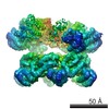
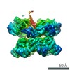
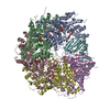
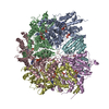
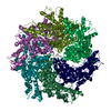
| Entry | Database: EMDB / ID: EMD-9698 | |||||||||
|---|---|---|---|---|---|---|---|---|---|---|
| Title | NSF-D1D2 part in the whole 20S complex | |||||||||
 Map data Map data | ||||||||||
 Sample Sample |
| |||||||||
 Keywords Keywords | membrane fusion / ATPase / HYDROLASE | |||||||||
| Function / homology |  Function and homology information Function and homology informationSNARE complex disassembly / ATP-dependent protein disaggregase activity / intra-Golgi vesicle-mediated transport / Golgi to plasma membrane protein transport / Golgi stack / syntaxin-1 binding / vesicle-fusing ATPase / positive regulation of receptor recycling / ionotropic glutamate receptor binding / SNARE binding ...SNARE complex disassembly / ATP-dependent protein disaggregase activity / intra-Golgi vesicle-mediated transport / Golgi to plasma membrane protein transport / Golgi stack / syntaxin-1 binding / vesicle-fusing ATPase / positive regulation of receptor recycling / ionotropic glutamate receptor binding / SNARE binding / PDZ domain binding / potassium ion transport / intracellular protein transport / positive regulation of protein catabolic process / midbody / protein kinase binding / protein-containing complex binding / ATP hydrolysis activity / ATP binding / metal ion binding / identical protein binding / plasma membrane / cytosol Similarity search - Function | |||||||||
| Biological species |   Cricetulus griseus (Chinese hamster) Cricetulus griseus (Chinese hamster) | |||||||||
| Method | single particle reconstruction / cryo EM / Resolution: 3.7 Å | |||||||||
 Authors Authors | Huang X / Sun S | |||||||||
 Citation Citation | Journal: Sci Adv / Year: 2019 Title: Mechanistic insights into the SNARE complex disassembly. Authors: Xuan Huang / Shan Sun / Xiaojing Wang / Fenghui Fan / Qiang Zhou / Shan Lu / Yong Cao / Qiu-Wen Wang / Meng-Qiu Dong / Jun Yao / Sen-Fang Sui /  Abstract: NSF (-ethylmaleimide-sensitive factor) and α-SNAP (α-soluble NSF attachment protein) bind to the SNARE (soluble NSF attachment protein receptor) complex, the minimum machinery to mediate membrane ...NSF (-ethylmaleimide-sensitive factor) and α-SNAP (α-soluble NSF attachment protein) bind to the SNARE (soluble NSF attachment protein receptor) complex, the minimum machinery to mediate membrane fusion, to form a 20S complex, which disassembles the SNARE complex for reuse. We report the cryo-EM structures of the α-SNAP-SNARE subcomplex and the NSF-D1D2 domain in the 20S complex at 3.9- and 3.7-Å resolutions, respectively. Combined with the biochemical and electrophysiological analyses, we find that α-SNAPs use R116 through electrostatic interactions and L197 through hydrophobic interactions to apply force mainly on two positions of the VAMP protein to execute disassembly process. Furthermore, we define the interaction between the amino terminus of the SNARE helical bundle and the pore loop of the NSF-D1 domain and demonstrate its essential role as a potential anchor for SNARE complex disassembly. Our studies provide a rotation model of α-SNAP-mediated disassembly of the SNARE complex. | |||||||||
| History |
|
- Structure visualization
| Movie |
Movie viewer |
|---|---|
| Structure viewer | EM map: SurfViewMolmilJmol/JSmol |
| Supplemental images |

- Downloads & links
-EMDB archive
| Map data | emd_9698.map.gz | 14.6 MB | EMDB map data format | |
|---|---|---|---|---|
| Header (meta data) | emd-9698-v30.xmlemd-9698.xml | 9.7 KB 9.7 KB | Display Display | EMDB header |
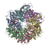
| Images | 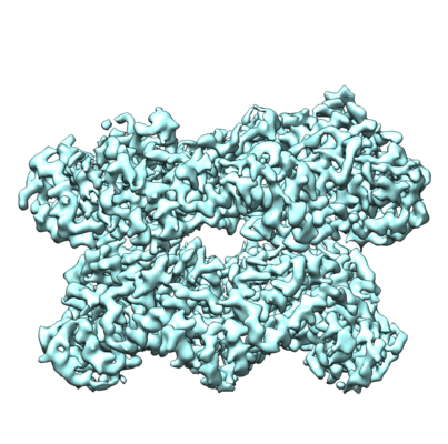 emd_9698.png emd_9698.png | 192.4 KB | ||
| Filedesc metadata | emd-9698.cif.gz | 5.7 KB | ||
| Archive directory |  http://ftp.pdbj.org/pub/emdb/structures/EMD-9698ftp://ftp.pdbj.org/pub/emdb/structures/EMD-9698 http://ftp.pdbj.org/pub/emdb/structures/EMD-9698ftp://ftp.pdbj.org/pub/emdb/structures/EMD-9698 | HTTPS FTP |
-Related structure data
| Related structure data |  6ip2MC  9697C  9723C 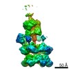 9724C  9725C  9726C 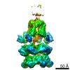 9727C  9728C  9729C  6ip1C M: atomic model generated by this map C: citing same article ( |
|---|---|
| Similar structure data |


-Links
| EMDB pages | EMDB (EBI/PDBe) / EMDataResource |
|---|---|
| Related items in Molecule of the Month |


-Map
| File | Download / File: emd_9698.map.gz / Format: CCP4 / Size: 15.6 MB / Type: IMAGE STORED AS FLOATING POINT NUMBER (4 BYTES) | ||||||||||||||||||||||||||||||||||||||||||||||||||||||||||||
|---|---|---|---|---|---|---|---|---|---|---|---|---|---|---|---|---|---|---|---|---|---|---|---|---|---|---|---|---|---|---|---|---|---|---|---|---|---|---|---|---|---|---|---|---|---|---|---|---|---|---|---|---|---|---|---|---|---|---|---|---|---|
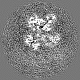
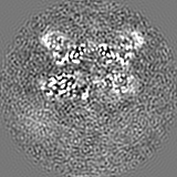
| Projections & slices | Image control
Images are generated by Spider. | ||||||||||||||||||||||||||||||||||||||||||||||||||||||||||||
| Voxel size | X=Y=Z: 1.30654 Å | ||||||||||||||||||||||||||||||||||||||||||||||||||||||||||||
| Density |
| ||||||||||||||||||||||||||||||||||||||||||||||||||||||||||||
| Symmetry | Space group: 1 | ||||||||||||||||||||||||||||||||||||||||||||||||||||||||||||
| Details | EMDB XML:
CCP4 map header:
| ||||||||||||||||||||||||||||||||||||||||||||||||||||||||||||
 Z (Sec.)
 Y (Row.)
 X (Col.)


-Supplemental data
- Sample components
-Entire : NSF-D1D2 part in the whole 20S complex
| Entire | Name: NSF-D1D2 part in the whole 20S complex |
|---|---|
| Components |
|
-Supramolecule #1: NSF-D1D2 part in the whole 20S complex
| Supramolecule | Name: NSF-D1D2 part in the whole 20S complex / type: complex / ID: 1 / Parent: 0 / Macromolecule list: #1 |
|---|---|
| Source (natural) | Organism: Cricetulus griseus (Chinese hamster) |
-Macromolecule #1: Vesicle-fusing ATPase
| Macromolecule | Name: Vesicle-fusing ATPase / type: protein_or_peptide / ID: 1 / Number of copies: 6 / Enantiomer: LEVO / EC number: vesicle-fusing ATPase |
|---|---|
| Source (natural) | Organism: Cricetulus griseus (Chinese hamster) |
| Molecular weight | Theoretical: 85.441133 KDa |
| Recombinant expression | Organism:  |
| Sequence | String: MRGSHHHHHH TDPLDVDSEP DVSAKMAGRS MQAARCPTDE LSLSNCAVVS EKDYQSGQHV IVRTSPNHKY IFTLRTHPSV VPGSVAFSL PQRKWAGLSI GQEIEVALYS FDKAKQCIGT MTIEIDFLQK KNIDSNPYDT DKMAAEFIQQ FNNQAFSVGQ Q LVFSFNDK ...String: MRGSHHHHHH TDPLDVDSEP DVSAKMAGRS MQAARCPTDE LSLSNCAVVS EKDYQSGQHV IVRTSPNHKY IFTLRTHPSV VPGSVAFSL PQRKWAGLSI GQEIEVALYS FDKAKQCIGT MTIEIDFLQK KNIDSNPYDT DKMAAEFIQQ FNNQAFSVGQ Q LVFSFNDK LFGLLVKDIE AVDPSILKGE PASGKRQKIE VGLVVGNSQV AFEKAENSSL NLIGKAKTKE NRQSIINPDW NF EKMGIGG LDKEFSDIFR RAFASRVFPP EIVEQMGCKH VKGILLYGPP GCGKTLLARQ IGKMLNAREP KVVNGPEILN KYV GESEAN IRKLFADAEE EQRRLGANSG LHIIIFDEID AICKQRGSMA GSTGVHDTVV NQLLSKIDGV EQLNNILVIG MTNR PDLID EALLRPGRLE VKMEIGLPDE KGRLQILHIH TARMRGHQLL SADVDIKELA VETKNFSGAE LEGLVRAAQS TAMNR HIKA STKVEVDMEK AESLQVTRGD FLASLENDIK PAFGTNQEDY ASYIMNGIIK WGDPVTRVLD DGELLVQQTK NSDRTP LVS VLLEGPPHSG KTALAAKIAE ESNFPFIKIC SPDKMIGFSE TAKCQAMKKI FDDAYKSQLS CVVVDDIERL LDYVPIG PR FSNLVLQALL VLLKKAPPQG RKLLIIGTTS RKDVLQEMEM LNAFSTTIHV PNIATGEQLL EALELLGNFK DKERTTIA Q QVKGKKVWIG IKKLLMLIEM SLQMDPEYRV RKFLALLREE GASPLDFD UniProtKB: Vesicle-fusing ATPase |
-Macromolecule #2: ADENOSINE-5'-TRIPHOSPHATE
| Macromolecule | Name: ADENOSINE-5'-TRIPHOSPHATE / type: ligand / ID: 2 / Number of copies: 12 / Formula: ATP |
|---|---|
| Molecular weight | Theoretical: 507.181 Da |
| Chemical component information |  ChemComp-ATP: |
-Experimental details
-Structure determination
| Method | cryo EM |
|---|---|
 Processing Processing | single particle reconstruction |
| Aggregation state | particle |
-Sample preparation
| Buffer | pH: 7.4 |
|---|---|
| Vitrification | Cryogen name: ETHANE |
- Electron microscopy
| Microscope | FEI TITAN KRIOS |
|---|---|
| Image recording | Film or detector model: GATAN K2 SUMMIT (4k x 4k) / Average electron dose: 50.0 e/Å2 |
| Electron beam | Acceleration voltage: 300 kV / Electron source:  FIELD EMISSION GUN FIELD EMISSION GUN |
| Electron optics | Illumination mode: FLOOD BEAM / Imaging mode: BRIGHT FIELD |
| Experimental equipment |  Model: Titan Krios / Image courtesy: FEI Company |
-Image processing
| Startup model | Type of model: EMDB MAP |
|---|---|
| Final reconstruction | Resolution.type: BY AUTHOR / Resolution: 3.7 Å / Resolution method: FSC 0.143 CUT-OFF / Number images used: 163942 |
| Initial angle assignment | Type: RANDOM ASSIGNMENT |
| Final angle assignment | Type: MAXIMUM LIKELIHOOD |
