- EMDB-11649: Akirin2 bound to the human 26S proteasome -
+
Open data
ID or keywords:
Loading...
-
Basic information
Entry
Database: EMDB / ID: EMD-11649
Title
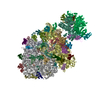
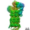
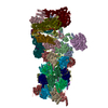
Akirin2 bound to the human 26S proteasome
Map data
Sample
Complex: Akirin2 bound to the human 26S proteasome
Function / homology
Function and homology information
proteasome localization / regulation of muscle cell differentiation / positive regulation of B cell activation / nuclear protein quality control by the ubiquitin-proteasome system / positive regulation of adaptive immune response / embryo development ending in birth or egg hatching / purine ribonucleoside triphosphate binding / positive regulation of innate immune response / Antigen processing: Ub, ATP-independent proteasomal degradation / sperm glycocalyx ...proteasome localization / regulation of muscle cell differentiation / positive regulation of B cell activation / nuclear protein quality control by the ubiquitin-proteasome system / positive regulation of adaptive immune response / embryo development ending in birth or egg hatching / purine ribonucleoside triphosphate binding / positive regulation of innate immune response / Antigen processing: Ub, ATP-independent proteasomal degradation / sperm glycocalyx / Regulation of ornithine decarboxylase (ODC) / Proteasome assembly / proteasome core complex / perinuclear theca / Cross-presentation of soluble exogenous antigens (endosomes) / Somitogenesis / myofibril / proteasomal ubiquitin-independent protein catabolic process / sperm head-tail coupling apparatus / immune system process / proteasome endopeptidase complex / NF-kappaB binding / proteasome core complex, beta-subunit complex / threonine-type endopeptidase activity / proteasome assembly / proteasome core complex, alpha-subunit complex / transcription repressor complex / : / ciliary tip / proteasome complex / sarcomere / Regulation of activated PAK-2p34 by proteasome mediated degradation / transcription coregulator activity / Autodegradation of Cdh1 by Cdh1:APC/C / APC/C:Cdc20 mediated degradation of Securin / Asymmetric localization of PCP proteins / centriole / Ubiquitin-dependent degradation of Cyclin D / sperm end piece / negative regulation of inflammatory response to antigenic stimulus / P-body / SCF-beta-TrCP mediated degradation of Emi1 / NIK-->noncanonical NF-kB signaling / AUF1 (hnRNP D0) binds and destabilizes mRNA / TNFR2 non-canonical NF-kB pathway / lipopolysaccharide binding / Assembly of the pre-replicative complex / Vpu mediated degradation of CD4 / Cdc20:Phospho-APC/C mediated degradation of Cyclin A / Dectin-1 mediated noncanonical NF-kB signaling / Degradation of DVL / Degradation of AXIN / Degradation of CRY and PER proteins / Hh mutants are degraded by ERAD / Activation of NF-kappaB in B cells / G2/M Checkpoints / Degradation of GLI1 by the proteasome / Hedgehog ligand biogenesis / Regulation of RUNX3 expression and activity / Autodegradation of the E3 ubiquitin ligase COP1 / Defective CFTR causes cystic fibrosis / cerebral cortex development / GSK3B and BTRC:CUL1-mediated-degradation of NFE2L2 / Negative regulation of NOTCH4 signaling / APC/C:Cdh1 mediated degradation of Cdc20 and other APC/C:Cdh1 targeted proteins in late mitosis/early G1 / Hedgehog 'on' state / Vif-mediated degradation of APOBEC3G / FBXL7 down-regulates AURKA during mitotic entry and in early mitosis / Degradation of GLI2 by the proteasome / GLI3 is processed to GLI3R by the proteasome / Ubiquitin-Mediated Degradation of Phosphorylated Cdc25A / MAPK6/MAPK4 signaling / positive regulation of interleukin-6 production / Degradation of CDH1 / Degradation of beta-catenin by the destruction complex / Oxygen-dependent proline hydroxylation of Hypoxia-inducible Factor Alpha / CDK-mediated phosphorylation and removal of Cdc6 / ABC-family protein mediated transport / CLEC7A (Dectin-1) signaling / SCF(Skp2)-mediated degradation of p27/p21 / FCERI mediated NF-kB activation / response to virus / nuclear matrix / Regulation of expression of SLITs and ROBOs / Regulation of PTEN stability and activity / Interleukin-1 signaling / protein import into nucleus / Orc1 removal from chromatin / Regulation of RAS by GAPs / Regulation of RUNX2 expression and activity / The role of GTSE1 in G2/M progression after G2 checkpoint / Separation of Sister Chromatids / UCH proteinases / KEAP1-NFE2L2 pathway / peptidase activity / Downstream TCR signaling / Antigen processing: Ubiquitination & Proteasome degradation / sperm principal piece / RUNX1 regulates transcription of genes involved in differentiation of HSCs / ER-Phagosome pathway Similarity search - Function
Journal: Nature / Year: 2021 Title: AKIRIN2 controls the nuclear import of proteasomes in vertebrates. Authors: Melanie de Almeida / Matthias Hinterndorfer / Hanna Brunner / Irina Grishkovskaya / Kashish Singh / Alexander Schleiffer / Julian Jude / Sumit Deswal / Robert Kalis / Milica Vunjak / Thomas ...Authors: Melanie de Almeida / Matthias Hinterndorfer / Hanna Brunner / Irina Grishkovskaya / Kashish Singh / Alexander Schleiffer / Julian Jude / Sumit Deswal / Robert Kalis / Milica Vunjak / Thomas Lendl / Richard Imre / Elisabeth Roitinger / Tobias Neumann / Susanne Kandolf / Michael Schutzbier / Karl Mechtler / Gijs A Versteeg / David Haselbach / Johannes Zuber / Abstract: Protein expression and turnover are controlled through a complex interplay of transcriptional, post-transcriptional and post-translational mechanisms to enable spatial and temporal regulation of ...Protein expression and turnover are controlled through a complex interplay of transcriptional, post-transcriptional and post-translational mechanisms to enable spatial and temporal regulation of cellular processes. To systematically elucidate such gene regulatory networks, we developed a CRISPR screening assay based on time-controlled Cas9 mutagenesis, intracellular immunostaining and fluorescence-activated cell sorting that enables the identification of regulatory factors independent of their effects on cellular fitness. We pioneered this approach by systematically probing the regulation of the transcription factor MYC, a master regulator of cell growth. Our screens uncover a highly conserved protein, AKIRIN2, that is essentially required for nuclear protein degradation. We found that AKIRIN2 forms homodimers that directly bind to fully assembled 20S proteasomes to mediate their nuclear import. During mitosis, proteasomes are excluded from condensing chromatin and re-imported into newly formed daughter nuclei in a highly dynamic, AKIRIN2-dependent process. Cells undergoing mitosis in the absence of AKIRIN2 become devoid of nuclear proteasomes, rapidly causing accumulation of MYC and other nuclear proteins. Collectively, our study reveals a dedicated pathway controlling the nuclear import of proteasomes in vertebrates and establishes a scalable approach to decipher regulators in essential cellular processes.
History
Deposition
Aug 21, 2020
-
Header (metadata) release
Sep 1, 2021
-
Map release
Sep 1, 2021
-
Update
Dec 1, 2021
-
Current status
Dec 1, 2021
Processing site: PDBe / Status: Released
-
Structure visualization
Movie
Surface view with section colored by density value
Entire : Akirin2 bound to the human 26S proteasome
Entire
Name: Akirin2 bound to the human 26S proteasome
Components
Complex: Akirin2 bound to the human 26S proteasome
-
Supramolecule #1: Akirin2 bound to the human 26S proteasome
Supramolecule
Name: Akirin2 bound to the human 26S proteasome / type: complex / ID: 1 / Parent: 0
Source (natural)
Organism: Homo sapiens (human)
Recombinant expression
Organism: Escherichia coli (E. coli)
-
Experimental details
-
Structure determination
Method
negative staining
Processing
single particle reconstruction
Aggregation state
particle
-
Sample preparation
Buffer
pH: 6.5
Staining
Type: NEGATIVE / Material: Uranyl Acetate
Grid
Model: Homemade / Support film - Material: CARBON / Support film - topology: CONTINUOUS / Support film - Film thickness: 2.0 nm / Pretreatment - Type: GLOW DISCHARGE / Pretreatment - Atmosphere: AIR
-
Electron microscopy
Microscope
FEI TECNAI 20
Image recording
Film or detector model: FEI EAGLE (2k x 2k) / Average electron dose: 60.0 e/Å2
Electron beam
Acceleration voltage: 200 kV / Electron source: LAB6
Electron optics
C2 aperture diameter: 100.0 µm / Illumination mode: FLOOD BEAM / Imaging mode: BRIGHT FIELD / Cs: 2.0 mm
-
Image processing
Final reconstruction
Applied symmetry - Point group: C1 (asymmetric) / Resolution.type: BY AUTHOR / Resolution: 21.0 Å / Resolution method: FSC 0.5 CUT-OFF / Software - Name: RELION (ver. 3.1) / Number images used: 36000
Initial angle assignment
Type: RANDOM ASSIGNMENT / Software - Name: RELION (ver. 3.1)
In the structure databanks used in Yorodumi, some data are registered as the other names, "COVID-19 virus" and "2019-nCoV". Here are the details of the virus and the list of structure data.
Jan 31, 2019. EMDB accession codes are about to change! (news from PDBe EMDB page)
EMDB accession codes are about to change! (news from PDBe EMDB page)
The allocation of 4 digits for EMDB accession codes will soon come to an end. Whilst these codes will remain in use, new EMDB accession codes will include an additional digit and will expand incrementally as the available range of codes is exhausted. The current 4-digit format prefixed with “EMD-” (i.e. EMD-XXXX) will advance to a 5-digit format (i.e. EMD-XXXXX), and so on. It is currently estimated that the 4-digit codes will be depleted around Spring 2019, at which point the 5-digit format will come into force.
The EM Navigator/Yorodumi systems omit the EMD- prefix.
Related info.:Q: What is EMD? / ID/Accession-code notation in Yorodumi/EM Navigator
Yorodumi is a browser for structure data from EMDB, PDB, SASBDB, etc.
This page is also the successor to EM Navigator detail page, and also detail information page/front-end page for Omokage search.
The word "yorodu" (or yorozu) is an old Japanese word meaning "ten thousand". "mi" (miru) is to see.
Related info.:EMDB / PDB / SASBDB / Comparison of 3 databanks / Yorodumi Search / Aug 31, 2016. New EM Navigator & Yorodumi / Yorodumi Papers / Jmol/JSmol / Function and homology information / Changes in new EM Navigator and Yorodumi
 Movie
Movie Controller


 Open data
 Basic information
 Map data
 Sample
 Function and homology information
 Homo sapiens (human)
 Authors
 Citation


 Structure visualization
 UCSF Chimera
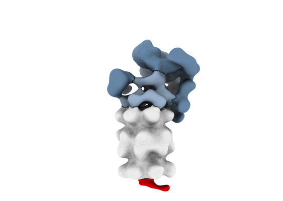
 Downloads & links
 emd_11649.png
 http://ftp.pdbj.org/pub/emdb/structures/EMD-11649


 Z (Sec.)
 Y (Row.)
 X (Col.)


 Sample components
 Processing
 Electron microscopy