 Movie
Movie Controller

[English] 日本語
 Yorodumi
- PDB-1j2q: 20S proteasome in complex with calpain-Inhibitor I from archaeogl... -
+ Open data

- Basic information
| Entry | Database: PDB / ID: 1j2q | ||||||
|---|---|---|---|---|---|---|---|
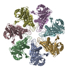
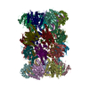
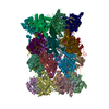
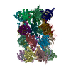
| Title | 20S proteasome in complex with calpain-Inhibitor I from archaeoglobus fulgidus | ||||||
 Components Components |
| ||||||
 Keywords Keywords | HYDROLASE / proteasome / ubiquitin / 20S / CP | ||||||
| Function / homology |  Function and homology information Function and homology informationproteasome endopeptidase complex / proteasome core complex, beta-subunit complex / threonine-type endopeptidase activity / proteasome core complex, alpha-subunit complex / proteasomal protein catabolic process / ubiquitin-dependent protein catabolic process / cytoplasm Similarity search - Function | ||||||
| Biological species |   Archaeoglobus fulgidus (archaea) Archaeoglobus fulgidus (archaea) | ||||||
| Method |  X-RAY DIFFRACTION / SYNCHROTRON / MOLECULAR REPLACEMENT / Resolution: 2.83 Å X-RAY DIFFRACTION / SYNCHROTRON / MOLECULAR REPLACEMENT / Resolution: 2.83 Å | ||||||
 Authors Authors | Groll, M. / Brandstetter, H. / Bartunik, H. / Bourenkow, G. / Huber, R. | ||||||
 Citation Citation | Journal: J.MOL.BIOL. / Year: 2003 Title: Investigations on the Maturation and Regulation of Archaebacterial Proteasomes Authors: Groll, M. / Brandstetter, H. / Bartunik, H. / Bourenkow, G. / Huber, R. | ||||||
| History |
|
- Structure visualization
| Structure viewer | Molecule: MolmilJmol/JSmol |
|---|
- Downloads & links
-Download
| PDBx/mmCIF format | 1j2q.cif.gz | 583.3 KB | Display | PDBx/mmCIF format |
|---|---|---|---|---|
| PDB format | pdb1j2q.ent.gz | 484.6 KB | Display | PDB format |
| PDBx/mmJSON format | 1j2q.json.gz | Tree view | PDBx/mmJSON format | |
| Others |  Other downloads Other downloads |
-Validation report
| Arichive directory | https://data.pdbj.org/pub/pdb/validation_reports/j2/1j2qftp://data.pdbj.org/pub/pdb/validation_reports/j2/1j2q | HTTPS FTP |
|---|
-Related structure data


-Links
 PDBj
PDBj


- Assembly
| Deposited unit | 
| ||||||||
|---|---|---|---|---|---|---|---|---|---|
| 1 | 
| ||||||||
| Unit cell |
| ||||||||
| Details | The second part of the bioogical assembly is generated by the two-fold axis: x, -y, -z+2/3 |
-Components
| #1: Protein | Mass: 26561.521 Da / Num. of mol.: 7 Source method: isolated from a genetically manipulated source Source: (gene. exp.) Archaeoglobus fulgidus (archaea) / Plasmid: prset6c / Production host:  References: UniProt: O29760, proteasome endopeptidase complex #2: Protein | Mass: 22132.283 Da / Num. of mol.: 7 Source method: isolated from a genetically manipulated source Source: (gene. exp.) Archaeoglobus fulgidus (archaea) / Plasmid: prset6c / Production host: References: UniProt: Q9P996, proteasome endopeptidase complex #3: Chemical | ChemComp-CIB /   Mass: 383.525 Da / Num. of mol.: 7 / Source method: obtained synthetically / Formula: C20H37N3O4 Mass: 383.525 Da / Num. of mol.: 7 / Source method: obtained synthetically / Formula: C20H37N3O4#4: Water | ChemComp-HOH / |  Mass: 18.015 Da / Num. of mol.: 105 / Source method: isolated from a natural source / Formula: H2O Mass: 18.015 Da / Num. of mol.: 105 / Source method: isolated from a natural source / Formula: H2OHas protein modification | N | |
|---|
-Experimental details
-Experiment
| Experiment | Method: X-RAY DIFFRACTION / Number of used crystals: 1 |
|---|
- Sample preparation
| Crystal | Density Matthews: 2.81 Å3/Da / Density % sol: 56.3 % | |||||||||||||||||||||||||||||||||||
|---|---|---|---|---|---|---|---|---|---|---|---|---|---|---|---|---|---|---|---|---|---|---|---|---|---|---|---|---|---|---|---|---|---|---|---|---|
| Crystal grow | Temperature: 298 K / Method: vapor diffusion, hanging drop / pH: 4.6 Details: 9% PEG 400, 80mM MgCl2, 100mM NaAc, pH 4.6, VAPOR DIFFUSION, HANGING DROP, temperature 298K | |||||||||||||||||||||||||||||||||||
| Crystal grow | *PLUS Temperature: 24 ℃ | |||||||||||||||||||||||||||||||||||
| Components of the solutions | *PLUS
|
-Data collection
| Diffraction | Mean temperature: 100 K |
|---|---|
| Diffraction source | Source: SYNCHROTRON / Site: MPG/DESY, HAMBURG  / Beamline: BW6 / Wavelength: 1.1 Å / Beamline: BW6 / Wavelength: 1.1 Å |
| Detector | Type: MARRESEARCH / Detector: CCD / Date: Oct 5, 2000 |
| Radiation | Monochromator: Si 111 CHANNEL / Protocol: SINGLE WAVELENGTH / Monochromatic (M) / Laue (L): M / Scattering type: x-ray |
| Radiation wavelength | Wavelength: 1.1 Å / Relative weight: 1 |
| Reflection | Resolution: 2.8→20 Å / Num. obs: 87322 / % possible obs: 94.9 % / Observed criterion σ(F): 2 / Observed criterion σ(I): 2 / Biso Wilson estimate: 46.1 Å2 |
| Reflection shell | Resolution: 2.83→2.88 Å / % possible all: 95.6 |
| Reflection | *PLUS Lowest resolution: 20 Å / Num. measured all: 1140521 / Rmerge(I) obs: 0.071 |
| Reflection shell | *PLUS Rmerge(I) obs: 0.399 |
- Processing
| Software |
| |||||||||||||||||||||||||
|---|---|---|---|---|---|---|---|---|---|---|---|---|---|---|---|---|---|---|---|---|---|---|---|---|---|---|
| Refinement | Method to determine structure: MOLECULAR REPLACEMENT / Resolution: 2.83→16.99 Å / Rfactor Rfree error: 0.004 / Data cutoff high absF: 3321738.1 / Data cutoff low absF: 0 / Isotropic thermal model: RESTRAINED / Cross valid method: THROUGHOUT / σ(F): 2 / Stereochemistry target values: Engh & Huber
| |||||||||||||||||||||||||
| Solvent computation | Solvent model: FLAT MODEL / Bsol: 28.0264 Å2 / ksol: 0.307215 e/Å3 | |||||||||||||||||||||||||
| Displacement parameters | Biso mean: 51.8 Å2
| |||||||||||||||||||||||||
| Refine analyze |
| |||||||||||||||||||||||||
| Refinement step | Cycle: LAST / Resolution: 2.83→16.99 Å
| |||||||||||||||||||||||||
| Refine LS restraints |
| |||||||||||||||||||||||||
| LS refinement shell | Resolution: 2.83→2.97 Å / Rfactor Rfree error: 0.017 / Total num. of bins used: 6
| |||||||||||||||||||||||||
| Xplor file |
| |||||||||||||||||||||||||
| Refinement | *PLUS Highest resolution: 2.8 Å | |||||||||||||||||||||||||
| Solvent computation | *PLUS | |||||||||||||||||||||||||
| Displacement parameters | *PLUS | |||||||||||||||||||||||||
| Refine LS restraints | *PLUS
|
