 Movie
Movie Controller


+ Open data

- Basic information
| Entry | Database: PDB / ID: 7nhe | ||||||
|---|---|---|---|---|---|---|---|
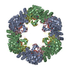
| Title | Crystal structure of Arabidopsis thaliana Pdx1K166R-I333 complex | ||||||
 Components Components | Pyridoxal 5'-phosphate synthase subunit PDX1.3 | ||||||
 Keywords Keywords | LYASE / VITAMIN B6 BIOSYTHESIS / PLP SYNTHASE / PDX1 | ||||||
| Function / homology |  Function and homology information Function and homology informationresponse to non-ionic osmotic stress / response to lipid hydroperoxide / chlorophyll metabolic process / amine-lyase activity / pyridoxal 5'-phosphate synthase (glutamine hydrolysing) / pyridoxal 5'-phosphate synthase (glutamine hydrolysing) activity / pyridoxal 5'-phosphate biosynthetic process / hyperosmotic salinity response / pyridoxine biosynthetic process / amino acid metabolic process ...response to non-ionic osmotic stress / response to lipid hydroperoxide / chlorophyll metabolic process / amine-lyase activity / pyridoxal 5'-phosphate synthase (glutamine hydrolysing) / pyridoxal 5'-phosphate synthase (glutamine hydrolysing) activity / pyridoxal 5'-phosphate biosynthetic process / hyperosmotic salinity response / pyridoxine biosynthetic process / amino acid metabolic process / response to UV-B / response to salt stress / endomembrane system / response to oxidative stress / protein heterodimerization activity / protein homodimerization activity / plasma membrane / cytosol Similarity search - Function | ||||||
| Biological species |  | ||||||
| Method |  X-RAY DIFFRACTION / SYNCHROTRON / FOURIER SYNTHESIS / Resolution: 2.23 Å X-RAY DIFFRACTION / SYNCHROTRON / FOURIER SYNTHESIS / Resolution: 2.23 Å | ||||||
 Authors Authors | Rodrigues, M.J. / Zhang, Y. / Bolton, R. / Evans, G. / Giri, N. / Royant, A. / Begley, T. / Ealick, S.E. / Tews, I. | ||||||
 Citation Citation | Journal: Rsc Chem Biol / Year: 2022 Title: Trapping and structural characterisation of a covalent intermediate in vitamin B 6 biosynthesis catalysed by the Pdx1 PLP synthase. Authors: Rodrigues, M.J. / Giri, N. / Royant, A. / Zhang, Y. / Bolton, R. / Evans, G. / Ealick, S.E. / Begley, T. / Tews, I. | ||||||
| History |
|
- Structure visualization
| Structure viewer | Molecule: MolmilJmol/JSmol |
|---|
- Downloads & links
-Download
| PDBx/mmCIF format | 7nhe.cif.gz | 420.6 KB | Display | PDBx/mmCIF format |
|---|---|---|---|---|
| PDB format | pdb7nhe.ent.gz | 346.1 KB | Display | PDB format |
| PDBx/mmJSON format | 7nhe.json.gz | Tree view | PDBx/mmJSON format | |
| Others |  Other downloads Other downloads |
-Validation report
| Arichive directory | https://data.pdbj.org/pub/pdb/validation_reports/nh/7nheftp://data.pdbj.org/pub/pdb/validation_reports/nh/7nhe | HTTPS FTP |
|---|
-Related structure data
| Related structure data | 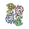 7nhfC  5lnuS S: Starting model for refinement C: citing same article ( |
|---|---|
| Similar structure data |


-Links
 PDBj
PDBj
- Assembly
| Deposited unit | 
| ||||||||||||||||||||
|---|---|---|---|---|---|---|---|---|---|---|---|---|---|---|---|---|---|---|---|---|---|
| 1 | 
| ||||||||||||||||||||
| Unit cell |
| ||||||||||||||||||||
| Noncrystallographic symmetry (NCS) | NCS oper:
|
-Components
| #1: Protein | Mass: 31210.072 Da / Num. of mol.: 4 / Mutation: K166R Source method: isolated from a genetically manipulated source Source: (gene. exp.)  References: UniProt: Q8L940, pyridoxal 5'-phosphate synthase (glutamine hydrolysing) #2: Chemical | ChemComp-PO4 /   Mass: 94.971 Da / Num. of mol.: 4 / Source method: isolated from a natural source / Formula: PO4 Mass: 94.971 Da / Num. of mol.: 4 / Source method: isolated from a natural source / Formula: PO4#3: Chemical | ChemComp-KPR / [(~{   Mass: 195.110 Da / Num. of mol.: 4 / Source method: isolated from a natural source / Formula: C5H10NO5P / Feature type: SUBJECT OF INVESTIGATION Mass: 195.110 Da / Num. of mol.: 4 / Source method: isolated from a natural source / Formula: C5H10NO5P / Feature type: SUBJECT OF INVESTIGATION#4: Water | ChemComp-HOH / |  Mass: 18.015 Da / Num. of mol.: 435 / Source method: isolated from a natural source / Formula: H2O Mass: 18.015 Da / Num. of mol.: 435 / Source method: isolated from a natural source / Formula: H2OHas ligand of interest | Y | Has protein modification | Y | |
|---|
-Experimental details
-Experiment
| Experiment | Method: X-RAY DIFFRACTION / Number of used crystals: 1 |
|---|
- Sample preparation
| Crystal | Density Matthews: 2.82 Å3/Da / Density % sol: 56.45 % |
|---|---|
| Crystal grow | Temperature: 291 K / Method: vapor diffusion, sitting drop / pH: 7 / Details: 28.1% PEG 1000 (w/v) and 100 mM HEPES pH 7.0 |
-Data collection
| Diffraction | Mean temperature: 100 K / Serial crystal experiment: N | ||||||||||||||||||||||||||||||
|---|---|---|---|---|---|---|---|---|---|---|---|---|---|---|---|---|---|---|---|---|---|---|---|---|---|---|---|---|---|---|---|
| Diffraction source | Source: SYNCHROTRON / Site: ESRF  / Beamline: ID23-1 / Wavelength: 0.980023 Å / Beamline: ID23-1 / Wavelength: 0.980023 Å | ||||||||||||||||||||||||||||||
| Detector | Type: DECTRIS PILATUS3 S 6M / Detector: PIXEL / Date: Jun 17, 2013 | ||||||||||||||||||||||||||||||
| Radiation | Protocol: SINGLE WAVELENGTH / Monochromatic (M) / Laue (L): M / Scattering type: x-ray | ||||||||||||||||||||||||||||||
| Radiation wavelength | Wavelength: 0.980023 Å / Relative weight: 1 | ||||||||||||||||||||||||||||||
| Reflection | Resolution: 2.23→92.23 Å / Num. obs: 66508 / % possible obs: 99.7 % / Redundancy: 3.9 % / CC1/2: 0.99 / Rmerge(I) obs: 0.104 / Rpim(I) all: 0.059 / Rrim(I) all: 0.12 / Net I/σ(I): 7.2 / Num. measured all: 260302 / Scaling rejects: 441 | ||||||||||||||||||||||||||||||
| Reflection shell | Diffraction-ID: 1
|
- Processing
| Software |
| |||||||||||||||||||||||||||||||||||||||||||||||||||||||||||||||||||||||||||||||||||||||||||||||||||||||||||||||||||||||||||||||||||||||||||||||||||||||||||||||||||||||||||||||
|---|---|---|---|---|---|---|---|---|---|---|---|---|---|---|---|---|---|---|---|---|---|---|---|---|---|---|---|---|---|---|---|---|---|---|---|---|---|---|---|---|---|---|---|---|---|---|---|---|---|---|---|---|---|---|---|---|---|---|---|---|---|---|---|---|---|---|---|---|---|---|---|---|---|---|---|---|---|---|---|---|---|---|---|---|---|---|---|---|---|---|---|---|---|---|---|---|---|---|---|---|---|---|---|---|---|---|---|---|---|---|---|---|---|---|---|---|---|---|---|---|---|---|---|---|---|---|---|---|---|---|---|---|---|---|---|---|---|---|---|---|---|---|---|---|---|---|---|---|---|---|---|---|---|---|---|---|---|---|---|---|---|---|---|---|---|---|---|---|---|---|---|---|---|---|---|---|
| Refinement | Method to determine structure: FOURIER SYNTHESIS Starting model: 5LNU Resolution: 2.23→92.23 Å / SU ML: 0.24 / Cross valid method: THROUGHOUT / σ(F): 1.94 / Phase error: 25.97 / Stereochemistry target values: ML
| |||||||||||||||||||||||||||||||||||||||||||||||||||||||||||||||||||||||||||||||||||||||||||||||||||||||||||||||||||||||||||||||||||||||||||||||||||||||||||||||||||||||||||||||
| Solvent computation | Shrinkage radii: 0.9 Å / VDW probe radii: 1.11 Å / Solvent model: FLAT BULK SOLVENT MODEL | |||||||||||||||||||||||||||||||||||||||||||||||||||||||||||||||||||||||||||||||||||||||||||||||||||||||||||||||||||||||||||||||||||||||||||||||||||||||||||||||||||||||||||||||
| Displacement parameters | Biso max: 123.53 Å2 / Biso mean: 48.4446 Å2 / Biso min: 28.44 Å2 | |||||||||||||||||||||||||||||||||||||||||||||||||||||||||||||||||||||||||||||||||||||||||||||||||||||||||||||||||||||||||||||||||||||||||||||||||||||||||||||||||||||||||||||||
| Refinement step | Cycle: final / Resolution: 2.23→92.23 Å
| |||||||||||||||||||||||||||||||||||||||||||||||||||||||||||||||||||||||||||||||||||||||||||||||||||||||||||||||||||||||||||||||||||||||||||||||||||||||||||||||||||||||||||||||
| LS refinement shell | Refine-ID: X-RAY DIFFRACTION / Rfactor Rfree error: 0 / Total num. of bins used: 24
| |||||||||||||||||||||||||||||||||||||||||||||||||||||||||||||||||||||||||||||||||||||||||||||||||||||||||||||||||||||||||||||||||||||||||||||||||||||||||||||||||||||||||||||||
| Refinement TLS params. | Method: refined / Refine-ID: X-RAY DIFFRACTION
| |||||||||||||||||||||||||||||||||||||||||||||||||||||||||||||||||||||||||||||||||||||||||||||||||||||||||||||||||||||||||||||||||||||||||||||||||||||||||||||||||||||||||||||||
| Refinement TLS group |
|
