 Movie
Movie Controller


+ Open data

- Basic information
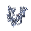
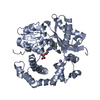
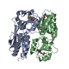
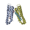
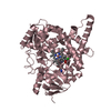
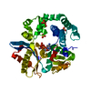
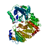
| Entry | Database: PDB / ID: 7nfq | |||||||||
|---|---|---|---|---|---|---|---|---|---|---|
| Title | Fujian capmidlink domain in complex with Nb8193 | |||||||||
 Components Components |
| |||||||||
 Keywords Keywords | VIRAL PROTEIN / Influenza polymerase / cap-binding domain / nanobody | |||||||||
| Function / homology |  Function and homology information Function and homology informationcap snatching / symbiont-mediated suppression of host mRNA transcription via inhibition of RNA polymerase II activity / host cell mitochondrion / 7-methylguanosine mRNA capping / virion component / symbiont-mediated suppression of host gene expression / viral RNA genome replication / RNA-directed RNA polymerase activity / DNA-templated transcription / host cell nucleus / RNA binding Similarity search - Function | |||||||||
| Biological species |   Influenza A virus Influenza A virus Camelidae mixed library (mammal) Camelidae mixed library (mammal) | |||||||||
| Method |  X-RAY DIFFRACTION / SYNCHROTRON / MOLECULAR REPLACEMENT / Resolution: 1.68 Å X-RAY DIFFRACTION / SYNCHROTRON / MOLECULAR REPLACEMENT / Resolution: 1.68 Å | |||||||||
 Authors Authors | Keown, J.R. / Grimes, J.M. / Fodor, E. | |||||||||
| Funding support |  United Kingdom, 2items United Kingdom, 2items
| |||||||||
 Citation Citation | Journal: Nat Commun / Year: 2022 Title: Mapping inhibitory sites on the RNA polymerase of the 1918 pandemic influenza virus using nanobodies. Authors: Jeremy R Keown / Zihan Zhu / Loïc Carrique / Haitian Fan / Alexander P Walker / Itziar Serna Martin / Els Pardon / Jan Steyaert / Ervin Fodor / Jonathan M Grimes /   Abstract: Influenza A viruses cause seasonal epidemics and global pandemics, representing a considerable burden to healthcare systems. Central to the replication cycle of influenza viruses is the viral RNA- ...Influenza A viruses cause seasonal epidemics and global pandemics, representing a considerable burden to healthcare systems. Central to the replication cycle of influenza viruses is the viral RNA-dependent RNA polymerase which transcribes and replicates the viral RNA genome. The polymerase undergoes conformational rearrangements and interacts with viral and host proteins to perform these functions. Here we determine the structure of the 1918 influenza virus polymerase in transcriptase and replicase conformations using cryo-electron microscopy (cryo-EM). We then structurally and functionally characterise the binding of single-domain nanobodies to the polymerase of the 1918 pandemic influenza virus. Combining these functional and structural data we identify five sites on the polymerase which are sensitive to inhibition by nanobodies. We propose that the binding of nanobodies at these sites either prevents the polymerase from assuming particular functional conformations or interactions with viral or host factors. The polymerase is highly conserved across the influenza A subtypes, suggesting these sites as effective targets for potential influenza antiviral development. | |||||||||
| History |
|
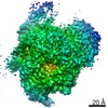
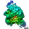
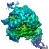
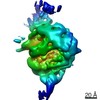
- Structure visualization
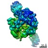
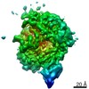
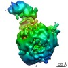
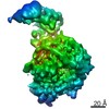
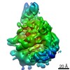
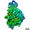
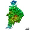
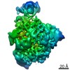
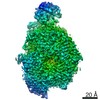
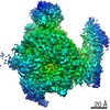
| Structure viewer | Molecule: MolmilJmol/JSmol |
|---|
- Downloads & links
-Download
| PDBx/mmCIF format | 7nfq.cif.gz | 566.5 KB | Display | PDBx/mmCIF format |
|---|---|---|---|---|
| PDB format | pdb7nfq.ent.gz | 387.7 KB | Display | PDB format |
| PDBx/mmJSON format | 7nfq.json.gz | Tree view | PDBx/mmJSON format | |
| Others |  Other downloads Other downloads |
-Validation report
| Arichive directory | https://data.pdbj.org/pub/pdb/validation_reports/nf/7nfqftp://data.pdbj.org/pub/pdb/validation_reports/nf/7nfq | HTTPS FTP |
|---|
-Related structure data
| Related structure data | 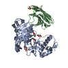 7nfrC 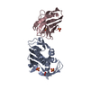 7nftC  7nhaC 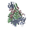 7nhcC 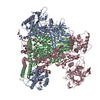 7nhxC  7ni0C  7nikC  7nilC 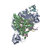 7nirC 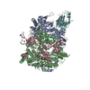 7nisC 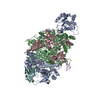 7nj3C 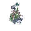 7nj4C  7nj5C 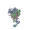 7nj7C 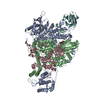 7nk1C 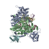 7nk2C  7nk4C 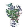 7nk6C  7nk8C 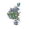 7nkaC 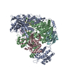 7nkcC  7nkiC  7nkrC  6s5vS S: Starting model for refinement C: citing same article ( |
|---|---|
| Similar structure data |


-Links
 PDBj
PDBj


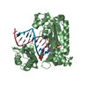
- Assembly
| Deposited unit | 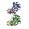
| ||||||||||||
|---|---|---|---|---|---|---|---|---|---|---|---|---|---|
| 1 | 
| ||||||||||||
| 2 | 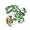
| ||||||||||||
| Unit cell |
|
-Components
| #1: Protein | Mass: 34249.168 Da / Num. of mol.: 2 Source method: isolated from a genetically manipulated source Source: (gene. exp.) Influenza A virus (A/duck/Fujian/13/2002(H5N1))Gene: PB2 / Production host:  #2: Antibody | Mass: 13983.466 Da / Num. of mol.: 2 Source method: isolated from a genetically manipulated source Source: (gene. exp.) Camelidae mixed library (mammal) / Production host: #3: Chemical | ChemComp-GOL /   Mass: 92.094 Da / Num. of mol.: 5 / Source method: obtained synthetically / Formula: C3H8O3 Mass: 92.094 Da / Num. of mol.: 5 / Source method: obtained synthetically / Formula: C3H8O3#4: Chemical | ChemComp-PEG / |   Mass: 106.120 Da / Num. of mol.: 1 / Source method: obtained synthetically / Formula: C4H10O3 Mass: 106.120 Da / Num. of mol.: 1 / Source method: obtained synthetically / Formula: C4H10O3#5: Water | ChemComp-HOH / |  Mass: 18.015 Da / Num. of mol.: 453 / Source method: isolated from a natural source / Formula: H2O Mass: 18.015 Da / Num. of mol.: 453 / Source method: isolated from a natural source / Formula: H2OHas ligand of interest | N | Has protein modification | Y | |
|---|
-Experimental details
-Experiment
| Experiment | Method: X-RAY DIFFRACTION / Number of used crystals: 1 |
|---|
- Sample preparation
| Crystal | Density Matthews: 2.29 Å3/Da / Density % sol: 46.32 % |
|---|---|
| Crystal grow | Temperature: 293 K / Method: vapor diffusion, sitting drop / Details: 20% w/v PEG3350 and 0.2 M (NH4)2 citrate |
-Data collection
| Diffraction | Mean temperature: 100 K / Serial crystal experiment: N |
|---|---|
| Diffraction source | Source: SYNCHROTRON / Site: Diamond / Beamline: I03 / Wavelength: 0.9762 Å |
| Detector | Type: DECTRIS EIGER2 X 16M / Detector: PIXEL / Date: May 14, 2020 |
| Radiation | Protocol: SINGLE WAVELENGTH / Monochromatic (M) / Laue (L): M / Scattering type: x-ray |
| Radiation wavelength | Wavelength: 0.9762 Å / Relative weight: 1 |
| Reflection | Resolution: 1.68→44.31 Å / Num. obs: 69149 / % possible obs: 70.31 % / Redundancy: 2 % / Biso Wilson estimate: 28.16 Å2 / CC1/2: 1 / Net I/σ(I): 23.17 |
| Reflection shell | Resolution: 1.68→1.743 Å / Num. unique obs: 767 / CC1/2: 0.292 |
- Processing
| Software |
| ||||||||||||||||||||||||||||||||||||||||||||||||||||||||||||||||||||||||||||||||||||||||||||||||||||||||||||||||||||||||||||||||||||||||||||||||||||||||||||||||||||||||||||||||||||||
|---|---|---|---|---|---|---|---|---|---|---|---|---|---|---|---|---|---|---|---|---|---|---|---|---|---|---|---|---|---|---|---|---|---|---|---|---|---|---|---|---|---|---|---|---|---|---|---|---|---|---|---|---|---|---|---|---|---|---|---|---|---|---|---|---|---|---|---|---|---|---|---|---|---|---|---|---|---|---|---|---|---|---|---|---|---|---|---|---|---|---|---|---|---|---|---|---|---|---|---|---|---|---|---|---|---|---|---|---|---|---|---|---|---|---|---|---|---|---|---|---|---|---|---|---|---|---|---|---|---|---|---|---|---|---|---|---|---|---|---|---|---|---|---|---|---|---|---|---|---|---|---|---|---|---|---|---|---|---|---|---|---|---|---|---|---|---|---|---|---|---|---|---|---|---|---|---|---|---|---|---|---|---|---|
| Refinement | Method to determine structure: MOLECULAR REPLACEMENT Starting model: 6S5V Resolution: 1.68→44.31 Å / SU ML: 0.1727 / Cross valid method: FREE R-VALUE / σ(F): 1.36 / Phase error: 28.9662 Stereochemistry target values: GeoStd + Monomer Library + CDL v1.2
| ||||||||||||||||||||||||||||||||||||||||||||||||||||||||||||||||||||||||||||||||||||||||||||||||||||||||||||||||||||||||||||||||||||||||||||||||||||||||||||||||||||||||||||||||||||||
| Solvent computation | Shrinkage radii: 0.9 Å / VDW probe radii: 1.11 Å / Solvent model: FLAT BULK SOLVENT MODEL | ||||||||||||||||||||||||||||||||||||||||||||||||||||||||||||||||||||||||||||||||||||||||||||||||||||||||||||||||||||||||||||||||||||||||||||||||||||||||||||||||||||||||||||||||||||||
| Displacement parameters | Biso mean: 35.39 Å2 | ||||||||||||||||||||||||||||||||||||||||||||||||||||||||||||||||||||||||||||||||||||||||||||||||||||||||||||||||||||||||||||||||||||||||||||||||||||||||||||||||||||||||||||||||||||||
| Refinement step | Cycle: LAST / Resolution: 1.68→44.31 Å
| ||||||||||||||||||||||||||||||||||||||||||||||||||||||||||||||||||||||||||||||||||||||||||||||||||||||||||||||||||||||||||||||||||||||||||||||||||||||||||||||||||||||||||||||||||||||
| Refine LS restraints |
| ||||||||||||||||||||||||||||||||||||||||||||||||||||||||||||||||||||||||||||||||||||||||||||||||||||||||||||||||||||||||||||||||||||||||||||||||||||||||||||||||||||||||||||||||||||||
| LS refinement shell |
| ||||||||||||||||||||||||||||||||||||||||||||||||||||||||||||||||||||||||||||||||||||||||||||||||||||||||||||||||||||||||||||||||||||||||||||||||||||||||||||||||||||||||||||||||||||||
| Refinement TLS params. | Method: refined / Origin x: 4.58878721433 Å / Origin y: 10.3509992209 Å / Origin z: 18.7829446686 Å
| ||||||||||||||||||||||||||||||||||||||||||||||||||||||||||||||||||||||||||||||||||||||||||||||||||||||||||||||||||||||||||||||||||||||||||||||||||||||||||||||||||||||||||||||||||||||
| Refinement TLS group | Selection details: all |
