 Movie
Movie Controller

[English] 日本語
 Yorodumi
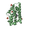
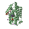
- PDB-7nfh: A heptameric barrel state of a de novo coiled-coil assembly: CC-T... -
+ Open data

- Basic information
| Entry | Database: PDB / ID: 7nfh | |||||||||
|---|---|---|---|---|---|---|---|---|---|---|
| Title | A heptameric barrel state of a de novo coiled-coil assembly: CC-Type2-(MaId)4. | |||||||||
 Components Components | CC-Type2-(MaId)4 | |||||||||
 Keywords Keywords | DE NOVO PROTEIN / Coiled coil / synthetic peptide / homomeric assembly | |||||||||
| Biological species | synthetic construct (others) | |||||||||
| Method |  X-RAY DIFFRACTION / SYNCHROTRON / MOLECULAR REPLACEMENT / Resolution: 1.62 Å X-RAY DIFFRACTION / SYNCHROTRON / MOLECULAR REPLACEMENT / Resolution: 1.62 Å | |||||||||
 Authors Authors | Rhys, G.G. / Dawson, W.M. / Brady, R.L. / Woolfson, D.N. | |||||||||
| Funding support | European Union,  United Kingdom, 2items United Kingdom, 2items
| |||||||||
 Citation Citation | Journal: Nat Commun / Year: 2023 Title: Differential sensing with arrays of de novo designed peptide assemblies. Authors: Dawson, W.M. / Shelley, K.L. / Fletcher, J.M. / Scott, D.A. / Lombardi, L. / Rhys, G.G. / LaGambina, T.J. / Obst, U. / Burton, A.J. / Cross, J.A. / Davies, G. / Martin, F.J.O. / Wiseman, F.J. ...Authors: Dawson, W.M. / Shelley, K.L. / Fletcher, J.M. / Scott, D.A. / Lombardi, L. / Rhys, G.G. / LaGambina, T.J. / Obst, U. / Burton, A.J. / Cross, J.A. / Davies, G. / Martin, F.J.O. / Wiseman, F.J. / Brady, R.L. / Tew, D. / Wood, C.W. / Woolfson, D.N. | |||||||||
| History |
|
- Structure visualization
| Structure viewer | Molecule:  MolmilJmol/JSmol MolmilJmol/JSmol |
|---|
- Downloads & links
-Download
| PDBx/mmCIF format | 7nfh.cif.gz | 105.2 KB | Display | PDBx/mmCIF format |
|---|---|---|---|---|
| PDB format | pdb7nfh.ent.gz | 85.7 KB | Display | PDB format |
| PDBx/mmJSON format | 7nfh.json.gz | Tree view | PDBx/mmJSON format | |
| Others |  Other downloads Other downloads |
-Validation report
| Arichive directory | https://data.pdbj.org/pub/pdb/validation_reports/nf/7nfhftp://data.pdbj.org/pub/pdb/validation_reports/nf/7nfh | HTTPS FTP |
|---|
-Related structure data
| Related structure data | 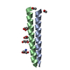 7nffC 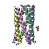 7nfgC 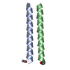 7nfiC 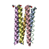 7nfjC 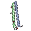 7nfkC 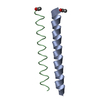 7nflC 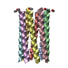 7nfmC  7nfnC 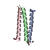 7nfoC 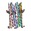 7nfpC  8a09C C: citing same article ( |
|---|---|
| Similar structure data |


-Links
 PDBj
PDBj
- Assembly
| Deposited unit | 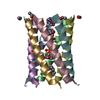
| |||||||||
|---|---|---|---|---|---|---|---|---|---|---|
| 1 |
| |||||||||
| Unit cell |
| |||||||||
| Components on special symmetry positions |
|
-Components
| #1: Protein/peptide | Mass: 3307.023 Da / Num. of mol.: 7 / Source method: obtained synthetically / Source: (synth.) synthetic construct (others) #2: Chemical | ChemComp-MES /   Mass: 195.237 Da / Num. of mol.: 5 / Source method: obtained synthetically / Formula: C6H13NO4S / Comment: pH buffer*YM Mass: 195.237 Da / Num. of mol.: 5 / Source method: obtained synthetically / Formula: C6H13NO4S / Comment: pH buffer*YM#3: Water | ChemComp-HOH / |  Mass: 18.015 Da / Num. of mol.: 94 / Source method: isolated from a natural source / Formula: H2O Mass: 18.015 Da / Num. of mol.: 94 / Source method: isolated from a natural source / Formula: H2OHas ligand of interest | N | Has protein modification | Y | |
|---|
-Experimental details
-Experiment
| Experiment | Method: X-RAY DIFFRACTION / Number of used crystals: 1 |
|---|
- Sample preparation
| Crystal | Density Matthews: 3.79 Å3/Da / Density % sol: 67.52 % |
|---|---|
| Crystal grow | Temperature: 292 K / Method: vapor diffusion, sitting drop / pH: 6.5 Details: After 1:1 dilution with the peptide solution, the resulting conditions were 250 mM ammonium sulfate and 50 mM 2-morpholinoethanesulfonic acid (MES). |
-Data collection
| Diffraction | Mean temperature: 100 K / Serial crystal experiment: N | |||||||||||||||||||||||||||
|---|---|---|---|---|---|---|---|---|---|---|---|---|---|---|---|---|---|---|---|---|---|---|---|---|---|---|---|---|
| Diffraction source | Source: SYNCHROTRON / Site: Diamond / Beamline: I04-1 / Wavelength: 0.9282 Å | |||||||||||||||||||||||||||
| Detector | Type: DECTRIS PILATUS 6M-F / Detector: PIXEL / Date: Feb 11, 2017 | |||||||||||||||||||||||||||
| Radiation | Protocol: SINGLE WAVELENGTH / Monochromatic (M) / Laue (L): M / Scattering type: x-ray | |||||||||||||||||||||||||||
| Radiation wavelength | Wavelength: 0.9282 Å / Relative weight: 1 | |||||||||||||||||||||||||||
| Reflection | Resolution: 1.62→55.94 Å / Num. obs: 45086 / % possible obs: 99.7 % / Redundancy: 19.4 % / Biso Wilson estimate: 25.77 Å2 / CC1/2: 1 / CC star: 1 / Rmerge(I) obs: 0.106 / Rpim(I) all: 0.025 / Rrim(I) all: 0.109 / Net I/σ(I): 16.4 | |||||||||||||||||||||||||||
| Reflection shell | Diffraction-ID: 1 / Redundancy: 17.4 %
|
- Processing
| Software |
| ||||||||||||||||||||||||||||||||||||||||||||||||||||||||||||
|---|---|---|---|---|---|---|---|---|---|---|---|---|---|---|---|---|---|---|---|---|---|---|---|---|---|---|---|---|---|---|---|---|---|---|---|---|---|---|---|---|---|---|---|---|---|---|---|---|---|---|---|---|---|---|---|---|---|---|---|---|---|
| Refinement | Method to determine structure: MOLECULAR REPLACEMENT Starting model: model helices Resolution: 1.62→55.94 Å / Cor.coef. Fo:Fc: 0.968 / Cor.coef. Fo:Fc free: 0.943 / SU B: 9.7 / SU ML: 0.119 / Cross valid method: THROUGHOUT / σ(F): 0 / ESU R: 0.085 / ESU R Free: 0.088 / Stereochemistry target values: MAXIMUM LIKELIHOOD Details: HYDROGENS HAVE BEEN ADDED IN THE RIDING POSITIONS U VALUES : REFINED INDIVIDUALLY
| ||||||||||||||||||||||||||||||||||||||||||||||||||||||||||||
| Solvent computation | Ion probe radii: 0.8 Å / Shrinkage radii: 0.8 Å / VDW probe radii: 1.2 Å / Solvent model: MASK | ||||||||||||||||||||||||||||||||||||||||||||||||||||||||||||
| Displacement parameters | Biso max: 108.16 Å2 / Biso mean: 33.413 Å2 / Biso min: 18.18 Å2
| ||||||||||||||||||||||||||||||||||||||||||||||||||||||||||||
| Refinement step | Cycle: final / Resolution: 1.62→55.94 Å
| ||||||||||||||||||||||||||||||||||||||||||||||||||||||||||||
| Refine LS restraints |
| ||||||||||||||||||||||||||||||||||||||||||||||||||||||||||||
| LS refinement shell | Resolution: 1.62→1.662 Å / Rfactor Rfree error: 0 / Total num. of bins used: 20
|
