 Movie
Movie Controller

[English] 日本語
 Yorodumi
- PDB-7nfj: A heptameric barrel state of a de novo coiled-coil assembly: CC-T... -
+ Open data

- Basic information
| Entry | Database: PDB / ID: 7nfj | |||||||||
|---|---|---|---|---|---|---|---|---|---|---|
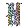
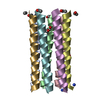
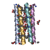
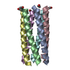
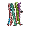
| Title | A heptameric barrel state of a de novo coiled-coil assembly: CC-Type2-(LaId)4-L28Y. | |||||||||
 Components Components | CC-Type2-(LaId)4-L28Y | |||||||||
 Keywords Keywords | DE NOVO PROTEIN / Coiled coil / synthetic peptide / homomeric assembly | |||||||||
| Biological species | synthetic construct (others) | |||||||||
| Method |  X-RAY DIFFRACTION / SYNCHROTRON / MOLECULAR REPLACEMENT / Resolution: 1.19 Å X-RAY DIFFRACTION / SYNCHROTRON / MOLECULAR REPLACEMENT / Resolution: 1.19 Å | |||||||||
 Authors Authors | Rhys, G.G. / Dawson, W.M. / Brady, R.L. / Woolfson, D.N. | |||||||||
| Funding support | European Union,  United Kingdom, 2items United Kingdom, 2items
| |||||||||
 Citation Citation | Journal: Nat Commun / Year: 2023 Title: Differential sensing with arrays of de novo designed peptide assemblies. Authors: Dawson, W.M. / Shelley, K.L. / Fletcher, J.M. / Scott, D.A. / Lombardi, L. / Rhys, G.G. / LaGambina, T.J. / Obst, U. / Burton, A.J. / Cross, J.A. / Davies, G. / Martin, F.J.O. / Wiseman, F.J. ...Authors: Dawson, W.M. / Shelley, K.L. / Fletcher, J.M. / Scott, D.A. / Lombardi, L. / Rhys, G.G. / LaGambina, T.J. / Obst, U. / Burton, A.J. / Cross, J.A. / Davies, G. / Martin, F.J.O. / Wiseman, F.J. / Brady, R.L. / Tew, D. / Wood, C.W. / Woolfson, D.N. | |||||||||
| History |
|
- Structure visualization
| Structure viewer | Molecule:  MolmilJmol/JSmol MolmilJmol/JSmol |
|---|
- Downloads & links
-Download
| PDBx/mmCIF format | 7nfj.cif.gz | 100.2 KB | Display | PDBx/mmCIF format |
|---|---|---|---|---|
| PDB format | pdb7nfj.ent.gz | 79.9 KB | Display | PDB format |
| PDBx/mmJSON format | 7nfj.json.gz | Tree view | PDBx/mmJSON format | |
| Others |  Other downloads Other downloads |
-Validation report
| Arichive directory | https://data.pdbj.org/pub/pdb/validation_reports/nf/7nfjftp://data.pdbj.org/pub/pdb/validation_reports/nf/7nfj | HTTPS FTP |
|---|
-Related structure data
| Related structure data | 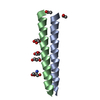 7nffC 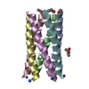 7nfgC 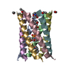 7nfhC 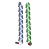 7nfiC 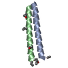 7nfkC 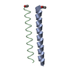 7nflC 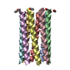 7nfmC  7nfnC 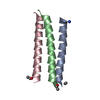 7nfoC 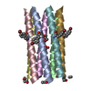 7nfpC 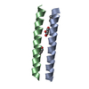 8a09C C: citing same article ( |
|---|---|
| Similar structure data |


-Links
 PDBj
PDBj
- Assembly
| Deposited unit | 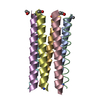
| |||||||||||||||||||||||||||||||||||||||||||||||||||||||||||||||||||||||||||||||||||||||||||||||||||||||||||||||||||||||||||||||||||||||||||||||||||||||||||||||||||||||||||||||||||||||||||||||||||||||||||||||||||||||||||||||||||||||||||||||||||||||||||||||||||||||||||||||||
|---|---|---|---|---|---|---|---|---|---|---|---|---|---|---|---|---|---|---|---|---|---|---|---|---|---|---|---|---|---|---|---|---|---|---|---|---|---|---|---|---|---|---|---|---|---|---|---|---|---|---|---|---|---|---|---|---|---|---|---|---|---|---|---|---|---|---|---|---|---|---|---|---|---|---|---|---|---|---|---|---|---|---|---|---|---|---|---|---|---|---|---|---|---|---|---|---|---|---|---|---|---|---|---|---|---|---|---|---|---|---|---|---|---|---|---|---|---|---|---|---|---|---|---|---|---|---|---|---|---|---|---|---|---|---|---|---|---|---|---|---|---|---|---|---|---|---|---|---|---|---|---|---|---|---|---|---|---|---|---|---|---|---|---|---|---|---|---|---|---|---|---|---|---|---|---|---|---|---|---|---|---|---|---|---|---|---|---|---|---|---|---|---|---|---|---|---|---|---|---|---|---|---|---|---|---|---|---|---|---|---|---|---|---|---|---|---|---|---|---|---|---|---|---|---|---|---|---|---|---|---|---|---|---|---|---|---|---|---|---|---|---|---|---|---|---|---|---|---|---|---|---|---|---|---|---|---|---|---|---|---|---|---|---|---|---|---|---|---|---|---|---|---|---|---|
| 1 |
| |||||||||||||||||||||||||||||||||||||||||||||||||||||||||||||||||||||||||||||||||||||||||||||||||||||||||||||||||||||||||||||||||||||||||||||||||||||||||||||||||||||||||||||||||||||||||||||||||||||||||||||||||||||||||||||||||||||||||||||||||||||||||||||||||||||||||||||||||
| Unit cell |
| |||||||||||||||||||||||||||||||||||||||||||||||||||||||||||||||||||||||||||||||||||||||||||||||||||||||||||||||||||||||||||||||||||||||||||||||||||||||||||||||||||||||||||||||||||||||||||||||||||||||||||||||||||||||||||||||||||||||||||||||||||||||||||||||||||||||||||||||||
| Noncrystallographic symmetry (NCS) | NCS domain:
NCS domain segments: Component-ID: _ / Beg auth comp-ID: GLY / Beg label comp-ID: GLY / End auth comp-ID: GLY / End label comp-ID: GLY / Refine code: _ / Auth seq-ID: 1 - 30 / Label seq-ID: 2 - 31
|