 Movie
Movie Controller

[English] 日本語
 Yorodumi
- PDB-7ncw: Crystal structure of oxidized glutaredoxin 2 from Chlamydomonas r... -
+ Open data

- Basic information
| Entry | Database: PDB / ID: 7ncw | ||||||
|---|---|---|---|---|---|---|---|
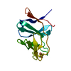
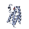
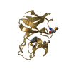
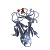
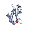
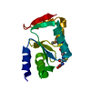
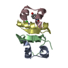
| Title | Crystal structure of oxidized glutaredoxin 2 from Chlamydomonas reinhardtii | ||||||
 Components Components | Glutaredoxin, CPYC type | ||||||
 Keywords Keywords | OXIDOREDUCTASE / Chlamydomonas reinhardtii glutaredoxin | ||||||
| Function / homology |  Function and homology information Function and homology informationglutathione disulfide oxidoreductase activity / cellular response to oxidative stress / cytoplasm Similarity search - Function | ||||||
| Biological species |   Chlamydomonas reinhardtii (plant) Chlamydomonas reinhardtii (plant) | ||||||
| Method |  X-RAY DIFFRACTION / MOLECULAR REPLACEMENT / Resolution: 2.4 Å X-RAY DIFFRACTION / MOLECULAR REPLACEMENT / Resolution: 2.4 Å | ||||||
 Authors Authors | Roret, T. / Didierjean, C. | ||||||
 Citation Citation | Journal: Antioxidants (Basel) / Year: 2021 Title: Atypical Iron-Sulfur Cluster Binding, Redox Activity and Structural Properties of Chlamydomonas reinhardtii Glutaredoxin 2. Authors: Roret, T. / Zhang, B. / Moseler, A. / Dhalleine, T. / Gao, X.H. / Couturier, J. / Lemaire, S.D. / Didierjean, C. / Johnson, M.K. / Rouhier, N. | ||||||
| History |
|
- Structure visualization
| Structure viewer | Molecule: MolmilJmol/JSmol |
|---|
- Downloads & links
-Download
| PDBx/mmCIF format | 7ncw.cif.gz | 65 KB | Display | PDBx/mmCIF format |
|---|---|---|---|---|
| PDB format | pdb7ncw.ent.gz | 39.1 KB | Display | PDB format |
| PDBx/mmJSON format | 7ncw.json.gz | Tree view | PDBx/mmJSON format | |
| Others |  Other downloads Other downloads |
-Validation report
| Arichive directory | https://data.pdbj.org/pub/pdb/validation_reports/nc/7ncwftp://data.pdbj.org/pub/pdb/validation_reports/nc/7ncw | HTTPS FTP |
|---|
-Related structure data
| Related structure data | 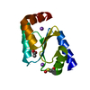 7ncvC  2flsS S: Starting model for refinement C: citing same article ( |
|---|---|
| Similar structure data |


-Links
 PDBj
PDBj- Assembly
| Deposited unit | 
| ||||||||||||
|---|---|---|---|---|---|---|---|---|---|---|---|---|---|
| 1 |
| ||||||||||||
| Unit cell |
| ||||||||||||
| Components on special symmetry positions |
|
-Components
| #1: Protein | Mass: 11356.931 Da / Num. of mol.: 1 Source method: isolated from a genetically manipulated source Details: Cys56 residue is oxidized into a sulfenic acid. / Source: (gene. exp.) Chlamydomonas reinhardtii (plant) / Gene: GRX2, CHLRE_12g550400v5, CHLREDRAFT_195611 / Production host:  |
|---|---|
| #2: Chemical | ChemComp-ACT /   Mass: 59.044 Da / Num. of mol.: 1 / Source method: obtained synthetically / Formula: C2H3O2 Mass: 59.044 Da / Num. of mol.: 1 / Source method: obtained synthetically / Formula: C2H3O2 |
| #3: Water | ChemComp-HOH /  Mass: 18.015 Da / Num. of mol.: 92 / Source method: isolated from a natural source / Formula: H2O Mass: 18.015 Da / Num. of mol.: 92 / Source method: isolated from a natural source / Formula: H2O |
| Has ligand of interest | N |
| Has protein modification | Y |
-Experimental details
-Experiment
| Experiment | Method: X-RAY DIFFRACTION / Number of used crystals: 1 |
|---|
- Sample preparation
| Crystal | Density Matthews: 2.73 Å3/Da / Density % sol: 54.92 % |
|---|---|
| Crystal grow | Temperature: 277 K / Method: microbatch Details: 30% (w/v) PEG 8000 0.1 M sodium cacodylate pH 6.5 0.2 M sodium acetate |
-Data collection
| Diffraction | Mean temperature: 100 K / Serial crystal experiment: N |
|---|---|
| Diffraction source | Source: SEALED TUBE / Type: OXFORD DIFFRACTION SUPERNOVA / Wavelength: 1.540562 Å |
| Detector | Type: AGILENT ATLAS CCD / Detector: CCD / Date: Jan 23, 2013 |
| Radiation | Protocol: SINGLE WAVELENGTH / Monochromatic (M) / Laue (L): M / Scattering type: x-ray |
| Radiation wavelength | Wavelength: 1.540562 Å / Relative weight: 1 |
| Reflection | Resolution: 2.4→14.7 Å / Num. obs: 5212 / % possible obs: 99.1 % / Redundancy: 6 % / Biso Wilson estimate: 21.36 Å2 / Rmerge(I) obs: 0.108 / Rpim(I) all: 0.046 / Rrim(I) all: 0.118 / Net I/σ(I): 12.3 |
| Reflection shell | Resolution: 2.4→2.53 Å / Rmerge(I) obs: 0.378 / Mean I/σ(I) obs: 3 / Num. unique obs: 723 / Rpim(I) all: 0.206 / Rrim(I) all: 0.435 |
- Processing
| Software |
| ||||||||||||||||||||||||||||||||||||||||
|---|---|---|---|---|---|---|---|---|---|---|---|---|---|---|---|---|---|---|---|---|---|---|---|---|---|---|---|---|---|---|---|---|---|---|---|---|---|---|---|---|---|
| Refinement | Method to determine structure: MOLECULAR REPLACEMENT Starting model: 2FLS Resolution: 2.4→14.3 Å / SU ML: 0.1968 / Cross valid method: FREE R-VALUE / σ(F): 1.34 / Phase error: 22.1177 Stereochemistry target values: GeoStd + Monomer Library + CDL v1.2
| ||||||||||||||||||||||||||||||||||||||||
| Solvent computation | Shrinkage radii: 0.9 Å / VDW probe radii: 1.11 Å / Solvent model: FLAT BULK SOLVENT MODEL | ||||||||||||||||||||||||||||||||||||||||
| Displacement parameters | Biso mean: 24.53 Å2 | ||||||||||||||||||||||||||||||||||||||||
| Refinement step | Cycle: LAST / Resolution: 2.4→14.3 Å
| ||||||||||||||||||||||||||||||||||||||||
| Refine LS restraints |
| ||||||||||||||||||||||||||||||||||||||||
| LS refinement shell |
| ||||||||||||||||||||||||||||||||||||||||
| Refinement TLS params. | Method: refined / Origin x: 3.57454576379 Å / Origin y: -19.7896406801 Å / Origin z: -9.96521343859 Å
| ||||||||||||||||||||||||||||||||||||||||
| Refinement TLS group | Selection details: (chain 'A' and resid 1 through 107) |
