 Movie
Movie Controller

[English] 日本語
 Yorodumi
- PDB-7mwy: Structure of the drosophila STING cyclic dinucleotide binding domain -
+ Open data

- Basic information
| Entry | Database: PDB / ID: 7mwy | ||||||
|---|---|---|---|---|---|---|---|
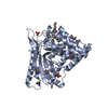
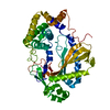
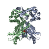
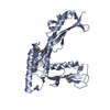
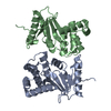
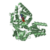
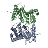
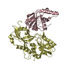
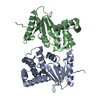
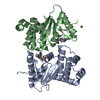
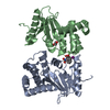
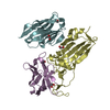
| Title | Structure of the drosophila STING cyclic dinucleotide binding domain | ||||||
 Components Components | STING | ||||||
 Keywords Keywords | IMMUNE SYSTEM / STING / cyclic dinucleotide receptor | ||||||
| Function / homology | Lysozyme - #40 / Lysozyme / Orthogonal Bundle / Mainly Alpha Function and homology information Function and homology information | ||||||
| Biological species |  | ||||||
| Method |  X-RAY DIFFRACTION / SYNCHROTRON / FOURIER SYNTHESIS / Resolution: 1.84 Å X-RAY DIFFRACTION / SYNCHROTRON / FOURIER SYNTHESIS / Resolution: 1.84 Å | ||||||
 Authors Authors | Slavik, K.M. / Ragucci, A.E. / Kranzusch, P.J. | ||||||
 Citation Citation | Journal: Nature / Year: 2021 Title: cGAS-like receptors sense RNA and control 3'2'-cGAMP signalling in Drosophila. Authors: Slavik, K.M. / Morehouse, B.R. / Ragucci, A.E. / Zhou, W. / Ai, X. / Chen, Y. / Li, L. / Wei, Z. / Bahre, H. / Konig, M. / Seifert, R. / Lee, A.S.Y. / Cai, H. / Imler, J.L. / Kranzusch, P.J. | ||||||
| History |
|
- Structure visualization
| Structure viewer | Molecule: MolmilJmol/JSmol |
|---|
- Downloads & links
-Download
| PDBx/mmCIF format | 7mwy.cif.gz | 195.3 KB | Display | PDBx/mmCIF format |
|---|---|---|---|---|
| PDB format | pdb7mwy.ent.gz | 129.5 KB | Display | PDB format |
| PDBx/mmJSON format | 7mwy.json.gz | Tree view | PDBx/mmJSON format | |
| Others |  Other downloads Other downloads |
-Validation report
| Arichive directory | https://data.pdbj.org/pub/pdb/validation_reports/mw/7mwyftp://data.pdbj.org/pub/pdb/validation_reports/mw/7mwy | HTTPS FTP |
|---|
-Related structure data

-Links
 PDBj
PDBj- Assembly
| Deposited unit | 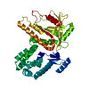
| ||||||||||||
|---|---|---|---|---|---|---|---|---|---|---|---|---|---|
| 1 |
| ||||||||||||
| Unit cell |
|
-Components
| #1: Protein | Mass: 40901.070 Da / Num. of mol.: 1 Source method: isolated from a genetically manipulated source Source: (gene. exp.)  |
|---|---|
| #2: Water | ChemComp-HOH /  Mass: 18.015 Da / Num. of mol.: 399 / Source method: isolated from a natural source / Formula: H2O Mass: 18.015 Da / Num. of mol.: 399 / Source method: isolated from a natural source / Formula: H2O |
-Experimental details
-Experiment
| Experiment | Method: X-RAY DIFFRACTION / Number of used crystals: 1 |
|---|
- Sample preparation
| Crystal | Density Matthews: 3.87 Å3/Da / Density % sol: 68.24 % |
|---|---|
| Crystal grow | Temperature: 291 K / Method: vapor diffusion, hanging drop / Details: 0.2 M sodium citrate, 0.1 M Tris-HCl, 22% PEG-3350 |
-Data collection
| Diffraction | Mean temperature: 80 K / Serial crystal experiment: N |
|---|---|
| Diffraction source | Source: SYNCHROTRON / Site: APS  / Beamline: 24-ID-E / Wavelength: 0.97918 Å / Beamline: 24-ID-E / Wavelength: 0.97918 Å |
| Detector | Type: DECTRIS EIGER X 16M / Detector: PIXEL / Date: Apr 1, 2021 |
| Radiation | Protocol: SINGLE WAVELENGTH / Monochromatic (M) / Laue (L): M / Scattering type: x-ray |
| Radiation wavelength | Wavelength: 0.97918 Å / Relative weight: 1 |
| Reflection | Resolution: 1.84→44.91 Å / Num. obs: 54801 / % possible obs: 99.7 % / Redundancy: 5.8 % / Biso Wilson estimate: 31.42 Å2 / CC1/2: 0.999 / Rpim(I) all: 0.035 / Net I/σ(I): 11.2 |
| Reflection shell | Resolution: 1.84→1.88 Å / Redundancy: 5.8 % / Mean I/σ(I) obs: 1.1 / Num. unique obs: 3340 / CC1/2: 0.435 / Rpim(I) all: 0.656 / % possible all: 98.6 |
- Processing
| Software |
| |||||||||||||||||||||||||||||||||||||||||||||||||||||||||||||||||||||||||||||||||||||||||||||||||||||||||
|---|---|---|---|---|---|---|---|---|---|---|---|---|---|---|---|---|---|---|---|---|---|---|---|---|---|---|---|---|---|---|---|---|---|---|---|---|---|---|---|---|---|---|---|---|---|---|---|---|---|---|---|---|---|---|---|---|---|---|---|---|---|---|---|---|---|---|---|---|---|---|---|---|---|---|---|---|---|---|---|---|---|---|---|---|---|---|---|---|---|---|---|---|---|---|---|---|---|---|---|---|---|---|---|---|---|---|
| Refinement | Method to determine structure: FOURIER SYNTHESIS Starting model: SAD-phased model of the selenomethionine derivative Resolution: 1.84→44.89 Å / SU ML: 0.2236 / Cross valid method: FREE R-VALUE / σ(F): 1.34 / Phase error: 20.5355 Stereochemistry target values: GeoStd + Monomer Library + CDL v1.2
| |||||||||||||||||||||||||||||||||||||||||||||||||||||||||||||||||||||||||||||||||||||||||||||||||||||||||
| Solvent computation | Shrinkage radii: 0.9 Å / VDW probe radii: 1.11 Å / Solvent model: FLAT BULK SOLVENT MODEL | |||||||||||||||||||||||||||||||||||||||||||||||||||||||||||||||||||||||||||||||||||||||||||||||||||||||||
| Displacement parameters | Biso mean: 40.32 Å2 | |||||||||||||||||||||||||||||||||||||||||||||||||||||||||||||||||||||||||||||||||||||||||||||||||||||||||
| Refinement step | Cycle: LAST / Resolution: 1.84→44.89 Å
| |||||||||||||||||||||||||||||||||||||||||||||||||||||||||||||||||||||||||||||||||||||||||||||||||||||||||
| Refine LS restraints |
| |||||||||||||||||||||||||||||||||||||||||||||||||||||||||||||||||||||||||||||||||||||||||||||||||||||||||
| LS refinement shell |
| |||||||||||||||||||||||||||||||||||||||||||||||||||||||||||||||||||||||||||||||||||||||||||||||||||||||||
| Refinement TLS params. | Method: refined / Refine-ID: X-RAY DIFFRACTION
| |||||||||||||||||||||||||||||||||||||||||||||||||||||||||||||||||||||||||||||||||||||||||||||||||||||||||
| Refinement TLS group | Refine-ID: X-RAY DIFFRACTION / Auth asym-ID: A / Label asym-ID: A
|
