Mass: 18.015 Da / Num. of mol.: 192 / Source method: isolated from a natural source / Formula: H2O
Has protein modification
Y
Sequence details
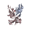
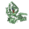
THE MISMATCH (RESIDUES 576-599) IS A WRONGLY ASSIGNMENT INTRON IN THE CHAETOMIUM THERMOPHILUM ...THE MISMATCH (RESIDUES 576-599) IS A WRONGLY ASSIGNMENT INTRON IN THE CHAETOMIUM THERMOPHILUM SEQUENCE. MULTIPLE SEQUENCE ALIGNMENTS WITH HOMOLOGUES FROM OTHER SPECIES SHOWED THAT THIS INTRON CORRESPONDS TO A PROTEIN SEQUENCE, WHICH IS PRESENT AND HIGHLY CONSERVED IN ALL OTHER SPECIES. IN ADDITION, ISOLATION OF THE GENE FROM A CHAETOMIUM THERMOPHILUM CDNA LIBRARY WITH APPROPRIATE PRIMERS AMPLIFIED THE GENE INCLUDING THE WRONGLY ASSIGNED INTRON.
-
Experimental details
-
Experiment
Experiment
Method: X-RAY DIFFRACTION / Number of used crystals: 1
-
Sample preparation
Crystal
Density Matthews: 2.38 Å3/Da / Density % sol: 48.42 %
Crystal grow
Temperature: 293 K / Method: vapor diffusion, sitting drop Details: 15 % PEG 8000, 0.5 M Li2SO4, VAPOR DIFFUSION, SITTING DROP, temperature 293K
-
Data collection
Diffraction
Mean temperature: 100 K
Diffraction source
Source: SYNCHROTRON / Site: PETRA III, EMBL c/o DESY / Beamline: P13 (MX1) / Wavelength: 0.82658 Å
In the structure databanks used in Yorodumi, some data are registered as the other names, "COVID-19 virus" and "2019-nCoV". Here are the details of the virus and the list of structure data.
Jan 31, 2019. EMDB accession codes are about to change! (news from PDBe EMDB page)
EMDB accession codes are about to change! (news from PDBe EMDB page)
The allocation of 4 digits for EMDB accession codes will soon come to an end. Whilst these codes will remain in use, new EMDB accession codes will include an additional digit and will expand incrementally as the available range of codes is exhausted. The current 4-digit format prefixed with “EMD-” (i.e. EMD-XXXX) will advance to a 5-digit format (i.e. EMD-XXXXX), and so on. It is currently estimated that the 4-digit codes will be depleted around Spring 2019, at which point the 5-digit format will come into force.
The EM Navigator/Yorodumi systems omit the EMD- prefix.
Related info.:Q: What is EMD? / ID/Accession-code notation in Yorodumi/EM Navigator
Yorodumi is a browser for structure data from EMDB, PDB, SASBDB, etc.
This page is also the successor to EM Navigator detail page, and also detail information page/front-end page for Omokage search.
The word "yorodu" (or yorozu) is an old Japanese word meaning "ten thousand". "mi" (miru) is to see.
Related info.:EMDB / PDB / SASBDB / Comparison of 3 databanks / Yorodumi Search / Aug 31, 2016. New EM Navigator & Yorodumi / Yorodumi Papers / Jmol/JSmol / Function and homology information / Changes in new EM Navigator and Yorodumi
 Movie
Movie Controller

 Yorodumi
 Open data
 Basic information
 Components
 Keywords
 Function and homology information
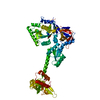
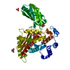
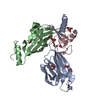
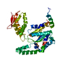
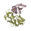
 Chaetomium thermophilum var. thermophilum (fungus)
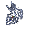
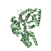
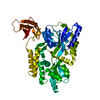
 X-RAY DIFFRACTION /
  Authors
 Citation
 Structure visualization
 Downloads & links
 Other downloads
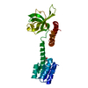


 PDBj
PDBj


 Assembly
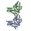


 Type: RNA linking / Mass: 443.201 Da / Num. of mol.: 2 / Source method: obtained synthetically / Formula: C10H15N5O11P2 / Comment: GDP, energy-carrying molecule*YM
 Mass: 24.305 Da / Num. of mol.: 2 / Source method: obtained synthetically / Formula: Mg
 Mass: 18.015 Da / Num. of mol.: 192 / Source method: isolated from a natural source / Formula: H2O
 Sample preparation
 / Beamline: P13 (MX1) / Wavelength: 0.82658 Å
 Processing