 Movie
Movie Controller

[English] 日本語
 Yorodumi
- PDB-7mwx: Structure of the core ectodomain of the hepatitis C virus envelop... -
+ Open data

- Basic information
| Entry | Database: PDB / ID: 7mwx | ||||||
|---|---|---|---|---|---|---|---|
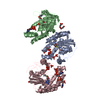
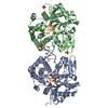
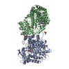
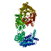
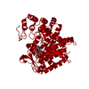
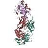
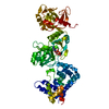
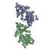
| Title | Structure of the core ectodomain of the hepatitis C virus envelope glycoprotein 2 with tamarin CD81 | ||||||
 Components Components |
| ||||||
 Keywords Keywords | VIRAL PROTEIN/IMMUNE SYSTEM / Glycoprotein / Receptor / Viral protein / VIRAL PROTEIN-IMMUNE SYSTEM complex | ||||||
| Function / homology |  Function and homology information Function and homology informationmacrophage fusion / CD4-positive, alpha-beta T cell costimulation / positive regulation of B cell receptor signaling pathway / osteoclast fusion / myoblast fusion involved in skeletal muscle regeneration / positive regulation of T cell activation via T cell receptor contact with antigen bound to MHC molecule on antigen presenting cell / positive regulation of inflammatory response to antigenic stimulus / regulation of macrophage migration / positive regulation of B cell activation / immunological synapse formation ...macrophage fusion / CD4-positive, alpha-beta T cell costimulation / positive regulation of B cell receptor signaling pathway / osteoclast fusion / myoblast fusion involved in skeletal muscle regeneration / positive regulation of T cell activation via T cell receptor contact with antigen bound to MHC molecule on antigen presenting cell / positive regulation of inflammatory response to antigenic stimulus / regulation of macrophage migration / positive regulation of B cell activation / immunological synapse formation / tetraspanin-enriched microdomain / positive regulation of T-helper 2 cell cytokine production / positive regulation of protein exit from endoplasmic reticulum / humoral immune response mediated by circulating immunoglobulin / positive regulation of CD4-positive, alpha-beta T cell proliferation / host cell mitochondrial membrane / host cell lipid droplet / cholesterol binding / symbiont-mediated transformation of host cell / symbiont-mediated suppression of host TRAF-mediated signal transduction / symbiont-mediated perturbation of host cell cycle G1/S transition checkpoint / positive regulation of T cell receptor signaling pathway / immunological synapse / cellular response to low-density lipoprotein particle stimulus / positive regulation of receptor clustering / symbiont-mediated suppression of host JAK-STAT cascade via inhibition of STAT1 activity / symbiont-mediated suppression of host cytoplasmic pattern recognition receptor signaling pathway via inhibition of MAVS activity / protein localization to plasma membrane / receptor internalization / integrin binding / ribonucleoside triphosphate phosphatase activity / channel activity / viral nucleocapsid / monoatomic ion transmembrane transport / clathrin-dependent endocytosis of virus by host cell / basolateral plasma membrane / RNA helicase activity / positive regulation of MAPK cascade / host cell perinuclear region of cytoplasm / host cell endoplasmic reticulum membrane / symbiont-mediated suppression of host type I interferon-mediated signaling pathway / ribonucleoprotein complex / symbiont-mediated activation of host autophagy / serine-type endopeptidase activity / cysteine-type endopeptidase activity / viral RNA genome replication / RNA-directed RNA polymerase activity / fusion of virus membrane with host endosome membrane / viral envelope / virion attachment to host cell / host cell nucleus / host cell plasma membrane / virion membrane / structural molecule activity / proteolysis / RNA binding / zinc ion binding / ATP binding / membrane / plasma membrane Similarity search - Function | ||||||
| Biological species |  Hepacivirus C Hepacivirus C Saguinus oedipus (cotton-top tamarin) Saguinus oedipus (cotton-top tamarin) | ||||||
| Method |  X-RAY DIFFRACTION / SYNCHROTRON / MOLECULAR REPLACEMENT / Resolution: 3.32 Å X-RAY DIFFRACTION / SYNCHROTRON / MOLECULAR REPLACEMENT / Resolution: 3.32 Å | ||||||
 Authors Authors | Kumar, A. / Hossain, R.A. / Yost, S.A. / Bu, W. / Wang, Y. / Dearborn, A.D. / Grakoui, A. / Cohen, J.I. / Marcotrigiano, J. | ||||||
| Funding support |  United States, 1items United States, 1items
| ||||||
 Citation Citation | Journal: Nature / Year: 2021 Title: Structural insights into hepatitis C virus receptor binding and entry. Authors: Kumar, A. / Hossain, R.A. / Yost, S.A. / Bu, W. / Wang, Y. / Dearborn, A.D. / Grakoui, A. / Cohen, J.I. / Marcotrigiano, J. | ||||||
| History |
|
- Structure visualization
| Structure viewer | Molecule: MolmilJmol/JSmol |
|---|
- Downloads & links
-Download
| PDBx/mmCIF format | 7mwx.cif.gz | 551.8 KB | Display | PDBx/mmCIF format |
|---|---|---|---|---|
| PDB format | pdb7mwx.ent.gz | 454.1 KB | Display | PDB format |
| PDBx/mmJSON format | 7mwx.json.gz | Tree view | PDBx/mmJSON format | |
| Others |  Other downloads Other downloads |
-Validation report
| Arichive directory | https://data.pdbj.org/pub/pdb/validation_reports/mw/7mwxftp://data.pdbj.org/pub/pdb/validation_reports/mw/7mwx | HTTPS FTP |
|---|
-Related structure data
| Related structure data |  7mwsC  7mwwC  4webS S: Starting model for refinement C: citing same article ( |
|---|---|
| Similar structure data |


-Links
 PDBj
PDBj


- Assembly
| Deposited unit | 
| ||||||||
|---|---|---|---|---|---|---|---|---|---|
| 1 | 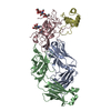
| ||||||||
| 2 | 
| ||||||||
| Unit cell |
|
-Components
-Protein , 2 types, 4 molecules CGDH
| #1: Protein | Mass: 29314.105 Da / Num. of mol.: 2 / Fragment: DeltaHVR1 Source method: isolated from a genetically manipulated source Details: J6 genotype / Source: (gene. exp.) Hepacivirus C / Cell line (production host): HEK293 gnti- cells / Production host:  Homo sapiens (human) Homo sapiens (human)References: UniProt: A0A2I6PIY1, RNA-directed RNA polymerase, hepacivirin, nucleoside-triphosphate phosphatase, RNA helicase #4: Protein | Mass: 10141.375 Da / Num. of mol.: 2 / Fragment: second extracellular domain Source method: isolated from a genetically manipulated source Source: (gene. exp.) Saguinus oedipus (cotton-top tamarin) / Gene: CD81 / Cell line (production host): HEK293 GNTI- cells / Production host: Homo sapiens (human) / References: UniProt: Q9N0J9 |
|---|
-Antibody , 2 types, 4 molecules AEBF
| #2: Antibody | Mass: 23207.604 Da / Num. of mol.: 2 Source method: isolated from a genetically manipulated source Source: (gene. exp.) Homo sapiens (human)#3: Antibody | Mass: 24111.613 Da / Num. of mol.: 2 Source method: isolated from a genetically manipulated source Source: (gene. exp.) Homo sapiens (human) |
|---|
-Sugars , 3 types, 5 molecules 
| #5: Polysaccharide | beta-D-mannopyranose-(1-4)-2-acetamido-2-deoxy-beta-D-glucopyranose-(1-4)-2-acetamido-2-deoxy-beta- ...beta-D-mannopyranose-(1-4)-2-acetamido-2-deoxy-beta-D-glucopyranose-(1-4)-2-acetamido-2-deoxy-beta-D-glucopyranose Source method: isolated from a genetically manipulated source |
|---|---|
| #6: Polysaccharide | 2-acetamido-2-deoxy-beta-D-glucopyranose-(1-4)-2-acetamido-2-deoxy-beta-D-glucopyranose Source method: isolated from a genetically manipulated source |
| #7: Sugar |  Type: D-saccharide, beta linking / Mass: 221.208 Da / Num. of mol.: 3 Type: D-saccharide, beta linking / Mass: 221.208 Da / Num. of mol.: 3Source method: isolated from a genetically manipulated source Formula: C8H15NO6 |
-Details
| Has ligand of interest | N |
|---|---|
| Has protein modification | Y |
-Experimental details
-Experiment
| Experiment | Method: X-RAY DIFFRACTION / Number of used crystals: 1 |
|---|
- Sample preparation
| Crystal | Density Matthews: 2.8 Å3/Da / Density % sol: 56.1 % |
|---|---|
| Crystal grow | Temperature: 277 K / Method: vapor diffusion, hanging drop / pH: 4.5 Details: 0.2M sodium acetate pH 4.5, 4% v/v tacsimate pH 4.0, 12% v/v PEG Smear Medium |
-Data collection
| Diffraction | Mean temperature: 100 K / Serial crystal experiment: N |
|---|---|
| Diffraction source | Source: SYNCHROTRON / Site: APS / Beamline: 22-ID / Wavelength: 0.979 Å |
| Detector | Type: DECTRIS EIGER2 XE 16M / Detector: PIXEL / Date: Dec 7, 2019 |
| Radiation | Protocol: SINGLE WAVELENGTH / Monochromatic (M) / Laue (L): M / Scattering type: x-ray |
| Radiation wavelength | Wavelength: 0.979 Å / Relative weight: 1 |
| Reflection | Resolution: 3.32→54.74 Å / Num. obs: 31609 / % possible obs: 99.5 % / Redundancy: 13.2 % / CC1/2: 0.99 / Net I/σ(I): 6.8 |
| Reflection shell | Resolution: 3.32→3.5 Å / Num. unique obs: 4559 / CC1/2: 0.39 |
- Processing
| Software |
| ||||||||||||||||||||||||||||||||||||||||||||||||||||||||||||||||||||||||||||||||||||
|---|---|---|---|---|---|---|---|---|---|---|---|---|---|---|---|---|---|---|---|---|---|---|---|---|---|---|---|---|---|---|---|---|---|---|---|---|---|---|---|---|---|---|---|---|---|---|---|---|---|---|---|---|---|---|---|---|---|---|---|---|---|---|---|---|---|---|---|---|---|---|---|---|---|---|---|---|---|---|---|---|---|---|---|---|---|
| Refinement | Method to determine structure: MOLECULAR REPLACEMENT Starting model: 4WEB Resolution: 3.32→53.093 Å / SU ML: 0.5 / Cross valid method: FREE R-VALUE / σ(F): 1.89 / Phase error: 33.83 / Stereochemistry target values: ML
| ||||||||||||||||||||||||||||||||||||||||||||||||||||||||||||||||||||||||||||||||||||
| Solvent computation | Shrinkage radii: 0.9 Å / VDW probe radii: 1.11 Å / Solvent model: FLAT BULK SOLVENT MODEL | ||||||||||||||||||||||||||||||||||||||||||||||||||||||||||||||||||||||||||||||||||||
| Refinement step | Cycle: LAST / Resolution: 3.32→53.093 Å
| ||||||||||||||||||||||||||||||||||||||||||||||||||||||||||||||||||||||||||||||||||||
| Refine LS restraints |
| ||||||||||||||||||||||||||||||||||||||||||||||||||||||||||||||||||||||||||||||||||||
| LS refinement shell |
| ||||||||||||||||||||||||||||||||||||||||||||||||||||||||||||||||||||||||||||||||||||
| Refinement TLS params. | Method: refined / Origin x: -13.9078 Å / Origin y: 16.0699 Å / Origin z: -28.4282 Å
| ||||||||||||||||||||||||||||||||||||||||||||||||||||||||||||||||||||||||||||||||||||
| Refinement TLS group | Selection details: all |
