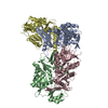
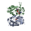
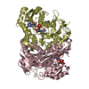
| Deposited unit | A: AMdnB protein
B: AMdnB protein
C: AMdnB protein
D: AMdnB protein
| Theoretical mass | Number of molelcules |
|---|
| Total (without water) | 165,142 | 4 |
|---|
| Polymers | 165,142 | 4 |
|---|
| Non-polymers | 0 | 0 |
|---|
| Water | 847 | 47 |
|---|
|
|---|
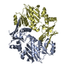
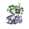
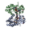
| 1 | A: AMdnB protein
D: AMdnB protein
| Theoretical mass | Number of molelcules |
|---|
| Total (without water) | 82,571 | 2 |
|---|
| Polymers | 82,571 | 2 |
|---|
| Non-polymers | 0 | 0 |
|---|
| Water | 36 | 2 |
|---|
| Type | Name | Symmetry operation | Number |
|---|
| identity operation | 1_555 | x,y,z | 1 |
| Buried area | 4860 Å2 |
|---|
| ΔGint | -32 kcal/mol |
|---|
| Surface area | 28110 Å2 |
|---|
| Method | PISA |
|---|
|
|---|
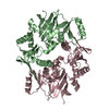
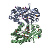
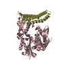
| 2 | B: AMdnB protein
C: AMdnB protein
| Theoretical mass | Number of molelcules |
|---|
| Total (without water) | 82,571 | 2 |
|---|
| Polymers | 82,571 | 2 |
|---|
| Non-polymers | 0 | 0 |
|---|
| Water | 36 | 2 |
|---|
| Type | Name | Symmetry operation | Number |
|---|
| identity operation | 1_555 | x,y,z | 1 |
| Buried area | 5100 Å2 |
|---|
| ΔGint | -28 kcal/mol |
|---|
| Surface area | 27840 Å2 |
|---|
| Method | PISA |
|---|
|
|---|
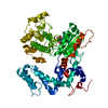
| Unit cell | | Length a, b, c (Å) | 61.479, 132.464, 83.010 |
|---|
| Angle α, β, γ (deg.) | 90.000, 91.005, 90.000 |
|---|
| Int Tables number | 4 |
|---|
| Space group name H-M | P1211 |
|---|
| Space group name Hall | P2yb |
|---|
| Symmetry operation | #1: x,y,z
#2: -x,y+1/2,-z |
|---|
|
|---|
| Noncrystallographic symmetry (NCS) | NCS domain:
NCS domain segments: Ens-ID: 1 | Dom-ID | Component-ID | Beg auth comp-ID | Beg label comp-ID | End auth comp-ID | End label comp-ID | Selection details | Auth asym-ID | Label asym-ID | Auth seq-ID | Label seq-ID |
|---|
| 1 | 1 | LEULEUSERSER(chain 'A' and (resid 3 through 178 or resid 197 through 237 or resid 252 through 326))AA| 3 - 177 | 37 - 211 | | 1 | 2 | LEULEUALAALA(chain 'A' and (resid 3 through 178 or resid 197 through 237 or resid 252 through 326))AA| 197 - 236 | 231 - 270 | | 1 | 3 | GLUGLUGLNGLN(chain 'A' and (resid 3 through 178 or resid 197 through 237 or resid 252 through 326))AA| 252 - 325 | 286 - 359 | | 2 | 1 | LEULEUSERSER(chain 'B' and (resid 3 through 178 or resid 197 through 237 or resid 252 through 326))BB| 3 - 177 | 37 - 211 | | 2 | 2 | LEULEUALAALA| (chain 'B' and (resid 3 through 178 or resid | | | | | | | | | | | | | | | | | | | | | | | | | | | | | | | | |
|
|---|
 Movie
Movie Controller

 Yorodumi
 Open data
 Basic information
 Components
 Keywords
 Function and homology information
 Nostoc sp. (bacteria)
 X-RAY DIFFRACTION /
  Authors
 Citation
 Structure visualization
 Downloads & links
 Other downloads
 PDBj
PDBj


 Assembly