 Movie
Movie Controller

[English] 日本語
 Yorodumi
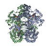
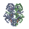
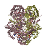
- PDB-7m32: Dihydropyrimidine Dehydrogenase (DPD) C671A Mutant Soaked with Ur... -
+ Open data

- Basic information
| Entry | Database: PDB / ID: 7m32 | ||||||
|---|---|---|---|---|---|---|---|
| Title | Dihydropyrimidine Dehydrogenase (DPD) C671A Mutant Soaked with Uracil and NADPH Anaerobically | ||||||
 Components Components | Dihydropyrimidine dehydrogenase [NADP(+)] | ||||||
 Keywords Keywords | FLAVOPROTEIN / Oxidoreductase / Dihydropyrimidine Dehydrogenase / DPD / mutant / uracil / flavin / soaking / ligand / FMN / FAD | ||||||
| Function / homology |  Function and homology information Function and homology informationdihydropyrimidine dehydrogenase (NADP+) / uracil binding / thymidine catabolic process / dihydropyrimidine dehydrogenase (NADP+) activity / beta-alanine biosynthetic process / uracil catabolic process / thymine catabolic process / NADP binding / FMN binding / flavin adenine dinucleotide binding ...dihydropyrimidine dehydrogenase (NADP+) / uracil binding / thymidine catabolic process / dihydropyrimidine dehydrogenase (NADP+) activity / beta-alanine biosynthetic process / uracil catabolic process / thymine catabolic process / NADP binding / FMN binding / flavin adenine dinucleotide binding / 4 iron, 4 sulfur cluster binding / protein homodimerization activity / metal ion binding / cytosol / cytoplasm Similarity search - Function | ||||||
| Biological species |  | ||||||
| Method |  X-RAY DIFFRACTION / SYNCHROTRON / MOLECULAR REPLACEMENT / Resolution: 1.82 Å X-RAY DIFFRACTION / SYNCHROTRON / MOLECULAR REPLACEMENT / Resolution: 1.82 Å | ||||||
 Authors Authors | Butrin, A. / Beaupre, B. / Forouzesh, D. / Liu, D. / Moran, G. | ||||||
 Citation Citation | Journal: Biochemistry / Year: 2021 Title: Perturbing the Movement of Hydrogens to Delineate and Assign Events in the Reductive Activation and Turnover of Porcine Dihydropyrimidine Dehydrogenase. Authors: Beaupre, B.A. / Forouzesh, D.C. / Butrin, A. / Liu, D. / Moran, G.R. | ||||||
| History |
|
- Structure visualization
| Structure viewer | Molecule: MolmilJmol/JSmol |
|---|
- Downloads & links
-Download
| PDBx/mmCIF format | 7m32.cif.gz | 846.7 KB | Display | PDBx/mmCIF format |
|---|---|---|---|---|
| PDB format | pdb7m32.ent.gz | 680.1 KB | Display | PDB format |
| PDBx/mmJSON format | 7m32.json.gz | Tree view | PDBx/mmJSON format | |
| Others |  Other downloads Other downloads |
-Validation report
| Arichive directory | https://data.pdbj.org/pub/pdb/validation_reports/m3/7m32ftp://data.pdbj.org/pub/pdb/validation_reports/m3/7m32 | HTTPS FTP |
|---|
-Related structure data
| Related structure data |  7m31C  1h7wS S: Starting model for refinement C: citing same article ( |
|---|---|
| Similar structure data |

-Links
 PDBj
PDBj


- Assembly
| Deposited unit | 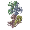
| ||||||||
|---|---|---|---|---|---|---|---|---|---|
| 1 | 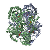
| ||||||||
| 2 | 
| ||||||||
| Unit cell |
|
-Components
-Protein , 1 types, 4 molecules ABCD
| #1: Protein | Mass: 111571.281 Da / Num. of mol.: 4 / Mutation: C671A Source method: isolated from a genetically manipulated source Source: (gene. exp.)  References: UniProt: Q28943, dihydropyrimidine dehydrogenase (NADP+) |
|---|
-Non-polymers , 8 types, 2663 molecules 


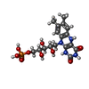


| #2: Chemical | ChemComp-SF4 /  Mass: 351.640 Da / Num. of mol.: 16 / Source method: obtained synthetically / Formula: Fe4S4 / Feature type: SUBJECT OF INVESTIGATION Mass: 351.640 Da / Num. of mol.: 16 / Source method: obtained synthetically / Formula: Fe4S4 / Feature type: SUBJECT OF INVESTIGATION#3: Chemical | ChemComp-FAD /  Mass: 785.550 Da / Num. of mol.: 4 / Source method: obtained synthetically / Formula: C27H33N9O15P2 / Feature type: SUBJECT OF INVESTIGATION / Comment: FAD*YM Mass: 785.550 Da / Num. of mol.: 4 / Source method: obtained synthetically / Formula: C27H33N9O15P2 / Feature type: SUBJECT OF INVESTIGATION / Comment: FAD*YM#4: Chemical | ChemComp-URA /  Mass: 112.087 Da / Num. of mol.: 4 / Source method: obtained synthetically / Formula: C4H4N2O2 / Feature type: SUBJECT OF INVESTIGATION Mass: 112.087 Da / Num. of mol.: 4 / Source method: obtained synthetically / Formula: C4H4N2O2 / Feature type: SUBJECT OF INVESTIGATION#5: Chemical | ChemComp-FNR /  Mass: 458.360 Da / Num. of mol.: 4 / Source method: obtained synthetically / Formula: C17H23N4O9P / Feature type: SUBJECT OF INVESTIGATION Mass: 458.360 Da / Num. of mol.: 4 / Source method: obtained synthetically / Formula: C17H23N4O9P / Feature type: SUBJECT OF INVESTIGATION#6: Chemical |  Type: L-peptide linking / Mass: 89.093 Da / Num. of mol.: 2 / Source method: obtained synthetically / Formula: C3H7NO2 / Feature type: SUBJECT OF INVESTIGATION Type: L-peptide linking / Mass: 89.093 Da / Num. of mol.: 2 / Source method: obtained synthetically / Formula: C3H7NO2 / Feature type: SUBJECT OF INVESTIGATION#7: Chemical |  Type: L-peptide linking / Mass: 131.173 Da / Num. of mol.: 2 / Source method: obtained synthetically / Formula: C6H13NO2 / Feature type: SUBJECT OF INVESTIGATION Type: L-peptide linking / Mass: 131.173 Da / Num. of mol.: 2 / Source method: obtained synthetically / Formula: C6H13NO2 / Feature type: SUBJECT OF INVESTIGATION#8: Chemical | ChemComp-PRO / |  Type: L-peptide linking / Mass: 115.130 Da / Num. of mol.: 1 / Source method: obtained synthetically / Formula: C5H9NO2 / Feature type: SUBJECT OF INVESTIGATION Type: L-peptide linking / Mass: 115.130 Da / Num. of mol.: 1 / Source method: obtained synthetically / Formula: C5H9NO2 / Feature type: SUBJECT OF INVESTIGATION#9: Water | ChemComp-HOH / | Mass: 18.015 Da / Num. of mol.: 2630 / Source method: isolated from a natural source / Formula: H2O |
|---|
-Details
| Has ligand of interest | Y |
|---|
-Experimental details
-Experiment
| Experiment | Method: X-RAY DIFFRACTION / Number of used crystals: 1 |
|---|
- Sample preparation
| Crystal | Density Matthews: 2.36 Å3/Da / Density % sol: 47.8 % |
|---|---|
| Crystal grow | Temperature: 293 K / Method: vapor diffusion, hanging drop Details: ~4.5 mg/mL (~40 uM) of C671A DPD in 25 mM HEPES, 2 mM DTT, 10% glycerol at pH 7.5 was prepared and diluted 1:1 with well solution containing 100 mM sodium citrate, 2 mM DTT, 15% PEG 6000 at ...Details: ~4.5 mg/mL (~40 uM) of C671A DPD in 25 mM HEPES, 2 mM DTT, 10% glycerol at pH 7.5 was prepared and diluted 1:1 with well solution containing 100 mM sodium citrate, 2 mM DTT, 15% PEG 6000 at pH 4.5 to 4.9. Diffraction quality crystals with elongated morphology were obtained by the hanging-drop vapor diffusion method. Incubation was carried out in the dark to eliminate photo-degradation the quasi-labile FMN cofactor. Crystals appeared after 16 hours as both single elongated rectangular hexahedron forms (200 x 50 x 50) or urchin-like clusters. Only the single crystal form were harvested and these were placed in a Plas-Labs 830 series glove box into which a Motic binocular microscope coupled to an Accuscope 1080 P HD camera was placed. Before being placed in the glove box, the well solutions of the selected crystals were made anaerobic with the addition of 10 mM dithionite and resealed with the cover slide. The glove box was then made anaerobic by flushing with pure nitrogen for approximately 10 minutes at which time the fractional dioxygen was 0.1 %, as indicated by a Forensics Detectors oxygen meter. Atmospheric dioxygen was measured throughout the soaking procedure and was held below 1%. C671A DPD crystals were soaked for a minimum of 20 minutes in 25 mM HEPES, 100 mM sodium citrate, 2 mM DTT, 100 uM NADPH, 100 uM uracil, 20% PEG 6000, 20% PEG 400, pH 7.5 prior to submersion in liquid nitrogen. Frozen crystals were then removed from the anaerobic environment and stored under liquid nitrogen. |
-Data collection
| Diffraction | Mean temperature: 100 K / Serial crystal experiment: N | ||||||||||||||||||||||||||||||
|---|---|---|---|---|---|---|---|---|---|---|---|---|---|---|---|---|---|---|---|---|---|---|---|---|---|---|---|---|---|---|---|
| Diffraction source | Source: SYNCHROTRON / Site: APS  / Beamline: 21-ID-D / Wavelength: 1.1272 Å / Beamline: 21-ID-D / Wavelength: 1.1272 Å | ||||||||||||||||||||||||||||||
| Detector | Type: DECTRIS EIGER X 9M / Detector: PIXEL / Date: Dec 9, 2020 | ||||||||||||||||||||||||||||||
| Radiation | Protocol: SINGLE WAVELENGTH / Monochromatic (M) / Laue (L): M / Scattering type: x-ray | ||||||||||||||||||||||||||||||
| Radiation wavelength | Wavelength: 1.1272 Å / Relative weight: 1 | ||||||||||||||||||||||||||||||
| Reflection | Resolution: 1.82→51.69 Å / Num. obs: 354924 / % possible obs: 96.5 % / Redundancy: 4.8 % / Biso Wilson estimate: 25.22 Å2 / CC1/2: 0.996 / Rmerge(I) obs: 0.147 / Rpim(I) all: 0.074 / Rrim(I) all: 0.165 / Net I/σ(I): 7.4 / Num. measured all: 1705022 / Scaling rejects: 41 | ||||||||||||||||||||||||||||||
| Reflection shell | Diffraction-ID: 1
|
- Processing
| Software |
| |||||||||||||||||||||||||||||||||||||||||||||||||||||||||||||||||||||||||||||||||||||||||||||||||||||||||||||||||||||||||||||||||||||||||||||||||||||||||||||||||||||||||||||||||||||||||||||||||||||||||||||||||||||||||
|---|---|---|---|---|---|---|---|---|---|---|---|---|---|---|---|---|---|---|---|---|---|---|---|---|---|---|---|---|---|---|---|---|---|---|---|---|---|---|---|---|---|---|---|---|---|---|---|---|---|---|---|---|---|---|---|---|---|---|---|---|---|---|---|---|---|---|---|---|---|---|---|---|---|---|---|---|---|---|---|---|---|---|---|---|---|---|---|---|---|---|---|---|---|---|---|---|---|---|---|---|---|---|---|---|---|---|---|---|---|---|---|---|---|---|---|---|---|---|---|---|---|---|---|---|---|---|---|---|---|---|---|---|---|---|---|---|---|---|---|---|---|---|---|---|---|---|---|---|---|---|---|---|---|---|---|---|---|---|---|---|---|---|---|---|---|---|---|---|---|---|---|---|---|---|---|---|---|---|---|---|---|---|---|---|---|---|---|---|---|---|---|---|---|---|---|---|---|---|---|---|---|---|---|---|---|---|---|---|---|---|---|---|---|---|---|---|---|---|
| Refinement | Method to determine structure: MOLECULAR REPLACEMENT Starting model: 1H7W Resolution: 1.82→47.26 Å / SU ML: 0.24 / Cross valid method: THROUGHOUT / σ(F): 1.34 / Phase error: 21.29 / Stereochemistry target values: ML
| |||||||||||||||||||||||||||||||||||||||||||||||||||||||||||||||||||||||||||||||||||||||||||||||||||||||||||||||||||||||||||||||||||||||||||||||||||||||||||||||||||||||||||||||||||||||||||||||||||||||||||||||||||||||||
| Solvent computation | Shrinkage radii: 0.9 Å / VDW probe radii: 1.11 Å / Solvent model: FLAT BULK SOLVENT MODEL | |||||||||||||||||||||||||||||||||||||||||||||||||||||||||||||||||||||||||||||||||||||||||||||||||||||||||||||||||||||||||||||||||||||||||||||||||||||||||||||||||||||||||||||||||||||||||||||||||||||||||||||||||||||||||
| Displacement parameters | Biso max: 131.41 Å2 / Biso mean: 34.5565 Å2 / Biso min: 13.88 Å2 | |||||||||||||||||||||||||||||||||||||||||||||||||||||||||||||||||||||||||||||||||||||||||||||||||||||||||||||||||||||||||||||||||||||||||||||||||||||||||||||||||||||||||||||||||||||||||||||||||||||||||||||||||||||||||
| Refinement step | Cycle: final / Resolution: 1.82→47.26 Å
| |||||||||||||||||||||||||||||||||||||||||||||||||||||||||||||||||||||||||||||||||||||||||||||||||||||||||||||||||||||||||||||||||||||||||||||||||||||||||||||||||||||||||||||||||||||||||||||||||||||||||||||||||||||||||
| LS refinement shell | Refine-ID: X-RAY DIFFRACTION / Rfactor Rfree error: 0 / Total num. of bins used: 30
|
