 Movie
Movie Controller


+ Open data

- Basic information
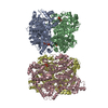
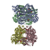
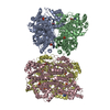
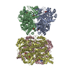
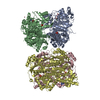
| Entry | Database: PDB / ID: 7lw1 | ||||||
|---|---|---|---|---|---|---|---|
| Title | Human phosphofructokinase-1 liver type bound to activator NA-11 | ||||||
 Components Components | ATP-dependent 6-phosphofructokinase, liver type | ||||||
 Keywords Keywords | TRANSFERASE / glycolysis / phosphofructokinase / liver type / activated | ||||||
| Function / homology |  Function and homology information Function and homology information6-phosphofructokinase complex / 6-phosphofructokinase / 6-phosphofructokinase activity / fructose binding / fructose-6-phosphate binding / fructose 1,6-bisphosphate metabolic process / fructose 6-phosphate metabolic process / Glycolysis / canonical glycolysis / response to glucose ...6-phosphofructokinase complex / 6-phosphofructokinase / 6-phosphofructokinase activity / fructose binding / fructose-6-phosphate binding / fructose 1,6-bisphosphate metabolic process / fructose 6-phosphate metabolic process / Glycolysis / canonical glycolysis / response to glucose / glycolytic process / negative regulation of insulin secretion / kinase binding / secretory granule lumen / ficolin-1-rich granule lumen / Neutrophil degranulation / extracellular exosome / extracellular region / ATP binding / membrane / metal ion binding / identical protein binding / cytosol Similarity search - Function | ||||||
| Biological species |  Homo sapiens (human) Homo sapiens (human) | ||||||
| Method | ELECTRON MICROSCOPY / single particle reconstruction / cryo EM / Resolution: 2.9 Å | ||||||
 Authors Authors | Lynch, E.M. / Kollman, J.M. / Webb, B. | ||||||
| Funding support |  United States, 1items United States, 1items
| ||||||
 Citation Citation | Journal: Cell / Year: 2021 Title: Selective activation of PFKL suppresses the phagocytic oxidative burst. Authors: Neri Amara / Madison P Cooper / Maria A Voronkova / Bradley A Webb / Eric M Lynch / Justin M Kollman / Taylur Ma / Kebing Yu / Zijuan Lai / Dewakar Sangaraju / Nobuhiko Kayagaki / Kim Newton ...Authors: Neri Amara / Madison P Cooper / Maria A Voronkova / Bradley A Webb / Eric M Lynch / Justin M Kollman / Taylur Ma / Kebing Yu / Zijuan Lai / Dewakar Sangaraju / Nobuhiko Kayagaki / Kim Newton / Matthew Bogyo / Steven T Staben / Vishva M Dixit / Abstract: In neutrophils, nicotinamide adenine dinucleotide phosphate (NADPH) generated via the pentose phosphate pathway fuels NADPH oxidase NOX2 to produce reactive oxygen species for killing invading ...In neutrophils, nicotinamide adenine dinucleotide phosphate (NADPH) generated via the pentose phosphate pathway fuels NADPH oxidase NOX2 to produce reactive oxygen species for killing invading pathogens. However, excessive NOX2 activity can exacerbate inflammation, as in acute respiratory distress syndrome (ARDS). Here, we use two unbiased chemical proteomic strategies to show that small-molecule LDC7559, or a more potent designed analog NA-11, inhibits the NOX2-dependent oxidative burst in neutrophils by activating the glycolytic enzyme phosphofructokinase-1 liver type (PFKL) and dampening flux through the pentose phosphate pathway. Accordingly, neutrophils treated with NA-11 had reduced NOX2-dependent outputs, including neutrophil cell death (NETosis) and tissue damage. A high-resolution structure of PFKL confirmed binding of NA-11 to the AMP/ADP allosteric activation site and explained why NA-11 failed to agonize phosphofructokinase-1 platelet type (PFKP) or muscle type (PFKM). Thus, NA-11 represents a tool for selective activation of PFKL, the main phosphofructokinase-1 isoform expressed in immune cells. | ||||||
| History |
|
- Structure visualization
| Movie |
Movie viewer |
|---|---|
| Structure viewer | Molecule: MolmilJmol/JSmol |
- Downloads & links
-Download
| PDBx/mmCIF format | 7lw1.cif.gz | 929.7 KB | Display | PDBx/mmCIF format |
|---|---|---|---|---|
| PDB format | pdb7lw1.ent.gz | 791 KB | Display | PDB format |
| PDBx/mmJSON format | 7lw1.json.gz | Tree view | PDBx/mmJSON format | |
| Others |  Other downloads Other downloads |
-Validation report
| Arichive directory | https://data.pdbj.org/pub/pdb/validation_reports/lw/7lw1ftp://data.pdbj.org/pub/pdb/validation_reports/lw/7lw1 | HTTPS FTP |
|---|
-Related structure data
| Related structure data | 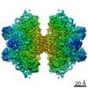 23544MC M: map data used to model this data C: citing same article ( |
|---|---|
| Similar structure data |
-Links
 PDBj
PDBj


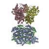
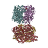
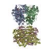
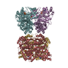
- Assembly
| Deposited unit | 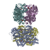
|
|---|---|
| 1 |
|
-Components
| #1: Protein | Mass: 85120.375 Da / Num. of mol.: 4 Source method: isolated from a genetically manipulated source Source: (gene. exp.) Homo sapiens (human) / Gene: PFKL / Production host:  unidentified baculovirus / References: UniProt: P17858, 6-phosphofructokinase unidentified baculovirus / References: UniProt: P17858, 6-phosphofructokinase#2: Chemical | ChemComp-YG1 /   Mass: 401.458 Da / Num. of mol.: 4 / Source method: obtained synthetically / Formula: C24H23N3O3 / Feature type: SUBJECT OF INVESTIGATION Mass: 401.458 Da / Num. of mol.: 4 / Source method: obtained synthetically / Formula: C24H23N3O3 / Feature type: SUBJECT OF INVESTIGATION#3: Sugar | ChemComp-FBP / 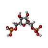  Type: D-saccharide, beta linking / Mass: 340.116 Da / Num. of mol.: 4 / Source method: obtained synthetically / Formula: C6H14O12P2 Type: D-saccharide, beta linking / Mass: 340.116 Da / Num. of mol.: 4 / Source method: obtained synthetically / Formula: C6H14O12P2#4: Sugar | ChemComp-F6P /   Type: D-saccharide, beta linking / Mass: 260.136 Da / Num. of mol.: 4 / Source method: obtained synthetically / Formula: C6H13O9P Type: D-saccharide, beta linking / Mass: 260.136 Da / Num. of mol.: 4 / Source method: obtained synthetically / Formula: C6H13O9P#5: Chemical | ChemComp-ADP /   Mass: 427.201 Da / Num. of mol.: 4 / Source method: obtained synthetically / Formula: C10H15N5O10P2 / Comment: ADP, energy-carrying molecule*YM Mass: 427.201 Da / Num. of mol.: 4 / Source method: obtained synthetically / Formula: C10H15N5O10P2 / Comment: ADP, energy-carrying molecule*YMHas ligand of interest | Y | |
|---|
-Experimental details
-Experiment
| Experiment | Method: ELECTRON MICROSCOPY |
|---|---|
| EM experiment | Aggregation state: PARTICLE / 3D reconstruction method: single particle reconstruction |
- Sample preparation
| Component | Name: Human phosphofructokinase-1 liver type bound to activator NA-11 Type: COMPLEX / Entity ID: #1 / Source: RECOMBINANT |
|---|---|
| Source (natural) | Organism: Homo sapiens (human) |
| Source (recombinant) | Organism: unidentified baculovirus |
| Buffer solution | pH: 7.5 |
| Specimen | Embedding applied: NO / Shadowing applied: NO / Staining applied: NO / Vitrification applied: YES |
| Specimen support | Grid material: COPPER / Grid mesh size: 400 divisions/in. / Grid type: C-flat-2/2 |
| Vitrification | Instrument: FEI VITROBOT MARK IV / Cryogen name: ETHANE / Humidity: 100 % |
- Electron microscopy imaging
| Experimental equipment |  Model: Titan Krios / Image courtesy: FEI Company |
|---|---|
| Microscopy | Model: FEI TITAN KRIOS |
| Electron gun | Electron source:  FIELD EMISSION GUN / Accelerating voltage: 300 kV / Illumination mode: FLOOD BEAM FIELD EMISSION GUN / Accelerating voltage: 300 kV / Illumination mode: FLOOD BEAM |
| Electron lens | Mode: BRIGHT FIELD / Nominal magnification: 130000 X / Nominal defocus max: 2800 nm / Nominal defocus min: 700 nm / Cs: 2.7 mm |
| Specimen holder | Cryogen: NITROGEN / Specimen holder model: FEI TITAN KRIOS AUTOGRID HOLDER |
| Image recording | Average exposure time: 10 sec. / Electron dose: 90 e/Å2 / Detector mode: SUPER-RESOLUTION / Film or detector model: GATAN K2 SUMMIT (4k x 4k) / Num. of grids imaged: 1 / Num. of real images: 2529 |
| Image scans | Width: 3710 / Height: 3838 / Movie frames/image: 50 / Used frames/image: 1-50 |
- Processing
| EM software |
| ||||||||||||||||||||||||||||||||
|---|---|---|---|---|---|---|---|---|---|---|---|---|---|---|---|---|---|---|---|---|---|---|---|---|---|---|---|---|---|---|---|---|---|
| CTF correction | Type: PHASE FLIPPING AND AMPLITUDE CORRECTION | ||||||||||||||||||||||||||||||||
| Particle selection | Num. of particles selected: 3000000 | ||||||||||||||||||||||||||||||||
| Symmetry | Point symmetry: D2 (2x2 fold dihedral) | ||||||||||||||||||||||||||||||||
| 3D reconstruction | Resolution: 2.9 Å / Resolution method: FSC 0.5 CUT-OFF / Num. of particles: 63296 / Symmetry type: POINT |
