 Movie
Movie Controller


+ Open data

- Basic information
| Entry | Database: PDB / ID: 3o8o | ||||||
|---|---|---|---|---|---|---|---|
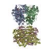
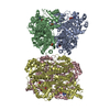
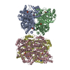
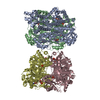
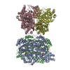
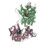
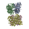
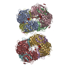
| Title | Structure of phosphofructokinase from Saccharomyces cerevisiae | ||||||
 Components Components |
| ||||||
 Keywords Keywords | TRANSFERASE / kinase | ||||||
| Function / homology |  Function and homology information Function and homology informationvacuolar proton-transporting V-type ATPase complex assembly / 6-phosphofructokinase complex / Glycolysis / 6-phosphofructokinase / 6-phosphofructokinase activity / fructose-6-phosphate binding / fructose 1,6-bisphosphate metabolic process / fructose 6-phosphate metabolic process / vacuolar acidification / canonical glycolysis ...vacuolar proton-transporting V-type ATPase complex assembly / 6-phosphofructokinase complex / Glycolysis / 6-phosphofructokinase / 6-phosphofructokinase activity / fructose-6-phosphate binding / fructose 1,6-bisphosphate metabolic process / fructose 6-phosphate metabolic process / vacuolar acidification / canonical glycolysis / Neutrophil degranulation / proton transmembrane transport / glycolytic process / regulation of intracellular pH / mitochondrial outer membrane / mRNA binding / mitochondrion / ATP binding / metal ion binding / cytoplasm Similarity search - Function | ||||||
| Biological species |  | ||||||
| Method |  X-RAY DIFFRACTION / SYNCHROTRON / MOLECULAR REPLACEMENT / Resolution: 2.9 Å X-RAY DIFFRACTION / SYNCHROTRON / MOLECULAR REPLACEMENT / Resolution: 2.9 Å | ||||||
 Authors Authors | Banaszak, K. / Mechin, I. / Kopperschlager, G. / Rypniewski, W. | ||||||
 Citation Citation | Journal: J.Mol.Biol. / Year: 2011 Title: The Crystal Structures of Eukaryotic Phosphofructokinases from Baker's Yeast and Rabbit Skeletal Muscle. Authors: Banaszak, K. / Mechin, I. / Obmolova, G. / Oldham, M. / Chang, S.H. / Ruiz, T. / Radermacher, M. / Kopperschlager, G. / Rypniewski, W. | ||||||
| History |
|
- Structure visualization
| Structure viewer | Molecule: MolmilJmol/JSmol |
|---|
- Downloads & links
-Download
| PDBx/mmCIF format | 3o8o.cif.gz | 1.1 MB | Display | PDBx/mmCIF format |
|---|---|---|---|---|
| PDB format | pdb3o8o.ent.gz | 911.6 KB | Display | PDB format |
| PDBx/mmJSON format | 3o8o.json.gz | Tree view | PDBx/mmJSON format | |
| Others |  Other downloads Other downloads |
-Validation report
| Arichive directory | https://data.pdbj.org/pub/pdb/validation_reports/o8/3o8oftp://data.pdbj.org/pub/pdb/validation_reports/o8/3o8o | HTTPS FTP |
|---|
-Related structure data
| Related structure data |  3o8lC  3o8nC  1pfkS S: Starting model for refinement C: citing same article ( |
|---|---|
| Similar structure data |
-Links
 PDBj
PDBj


- Assembly
| Deposited unit | 
| ||||||||
|---|---|---|---|---|---|---|---|---|---|
| 1 | 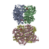
| ||||||||
| 2 | 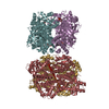
| ||||||||
| Unit cell |
|
-Components
| #1: Protein | Mass: 86002.547 Da / Num. of mol.: 4 Source method: isolated from a genetically manipulated source Source: (gene. exp.) Gene: PFK1, YGR240C, G8599 / Production host: #2: Protein | Mass: 83537.695 Da / Num. of mol.: 4 Source method: isolated from a genetically manipulated source Source: (gene. exp.) Gene: PFK2, YMR205C, YM8325.06C / Production host: #3: Sugar | ChemComp-F6P /   Type: D-saccharide, beta linking / Mass: 260.136 Da / Num. of mol.: 8 Type: D-saccharide, beta linking / Mass: 260.136 Da / Num. of mol.: 8Source method: isolated from a genetically manipulated source Formula: C6H13O9P #4: Sugar | ChemComp-FDP /   Type: D-saccharide, beta linking / Mass: 340.116 Da / Num. of mol.: 8 Type: D-saccharide, beta linking / Mass: 340.116 Da / Num. of mol.: 8Source method: isolated from a genetically manipulated source Formula: C6H14O12P2 |
|---|
-Experimental details
-Experiment
| Experiment | Method: X-RAY DIFFRACTION / Number of used crystals: 1 |
|---|
- Sample preparation
| Crystal | Density Matthews: 2.92 Å3/Da / Density % sol: 57.92 % |
|---|---|
| Crystal grow | Temperature: 277 K / Method: vapor diffusion, hanging drop / pH: 6 Details: 6-10% PEG4000, 0.2 mM sodium acetate, 0.1 M MES buffer pH 6.0, VAPOR DIFFUSION, HANGING DROP, temperature 277K |
-Data collection
| Diffraction | Mean temperature: 100 K |
|---|---|
| Diffraction source | Source: SYNCHROTRON / Site: EMBL/DESY, HAMBURG  / Beamline: BW7B / Wavelength: 1.1044 Å / Beamline: BW7B / Wavelength: 1.1044 Å |
| Detector | Type: MAR scanner 300 mm plate / Detector: IMAGE PLATE / Date: Apr 28, 1997 |
| Radiation | Monochromator: Si / Protocol: SINGLE WAVELENGTH / Monochromatic (M) / Laue (L): M / Scattering type: x-ray |
| Radiation wavelength | Wavelength: 1.1044 Å / Relative weight: 1 |
| Reflection | Resolution: 2.9→35 Å / Num. all: 172818 / Num. obs: 172818 / % possible obs: 98.5 % / Observed criterion σ(F): 0 / Observed criterion σ(I): 0 / Redundancy: 4 % / Rsym value: 0.075 / Net I/σ(I): 15.13 |
| Reflection shell | Resolution: 2.9→2.95 Å / Redundancy: 3 % / Mean I/σ(I) obs: 1.75 / Rsym value: 0.557 / % possible all: 98 |
- Processing
| Software |
| ||||||||||||||||||||||||||||||||||||||||||||||||||||||||||||
|---|---|---|---|---|---|---|---|---|---|---|---|---|---|---|---|---|---|---|---|---|---|---|---|---|---|---|---|---|---|---|---|---|---|---|---|---|---|---|---|---|---|---|---|---|---|---|---|---|---|---|---|---|---|---|---|---|---|---|---|---|---|
| Refinement | Method to determine structure: MOLECULAR REPLACEMENT Starting model: PDB ENTRY 1PFK Resolution: 2.9→35 Å / Isotropic thermal model: Isotropic / Cross valid method: THROUGHOUT / σ(F): 0 / σ(I): 0 / Stereochemistry target values: Engh & Huber
| ||||||||||||||||||||||||||||||||||||||||||||||||||||||||||||
| Refinement step | Cycle: LAST / Resolution: 2.9→35 Å
| ||||||||||||||||||||||||||||||||||||||||||||||||||||||||||||
| Refine LS restraints |
| ||||||||||||||||||||||||||||||||||||||||||||||||||||||||||||
| Xplor file |
|
