 Movie
Movie Controller

[English] 日本語
 Yorodumi
- PDB-4xyj: Crystal structure of human phosphofructokinase-1 in complex with ... -
+ Open data

- Basic information
| Entry | Database: PDB / ID: 4xyj | ||||||
|---|---|---|---|---|---|---|---|
| Title | Crystal structure of human phosphofructokinase-1 in complex with ATP and Mg, Northeast Structural Genomics Consortium Target HR9275 | ||||||
 Components Components | ATP-dependent 6-phosphofructokinase, platelet type | ||||||
 Keywords Keywords | TRANSFERASE / Structural Genomics / PSI-Biology / Protein Structure Initiative / Northeast Structural Genomics Consortium / NESG | ||||||
| Function / homology |  Function and homology information Function and homology information6-phosphofructokinase complex / 6-phosphofructokinase / 6-phosphofructokinase activity / fructose-6-phosphate binding / fructose 1,6-bisphosphate metabolic process / fructose 6-phosphate metabolic process / Glycolysis / canonical glycolysis / cellular response to leukemia inhibitory factor / cadherin binding ...6-phosphofructokinase complex / 6-phosphofructokinase / 6-phosphofructokinase activity / fructose-6-phosphate binding / fructose 1,6-bisphosphate metabolic process / fructose 6-phosphate metabolic process / Glycolysis / canonical glycolysis / cellular response to leukemia inhibitory factor / cadherin binding / protein-containing complex binding / extracellular exosome / ATP binding / membrane / metal ion binding / identical protein binding / nucleus / cytosol / cytoplasm Similarity search - Function | ||||||
| Biological species |  Homo sapiens (human) Homo sapiens (human) | ||||||
| Method |  X-RAY DIFFRACTION / SYNCHROTRON / MOLECULAR REPLACEMENT / Resolution: 3.1 Å X-RAY DIFFRACTION / SYNCHROTRON / MOLECULAR REPLACEMENT / Resolution: 3.1 Å | ||||||
 Authors Authors | Forouhar, F. / Webb, B.A. / Szu, F.-E. / Seetharaman, J. / Barber, D.L. / Tong, L. / Northeast Structural Genomics Consortium (NESG) | ||||||
 Citation Citation | Journal: Nature / Year: 2015 Title: Structures of human phosphofructokinase-1 and atomic basis of cancer-associated mutations. Authors: Webb, B.A. / Forouhar, F. / Szu, F.E. / Seetharaman, J. / Tong, L. / Barber, D.L. | ||||||
| History |
|
- Structure visualization
| Structure viewer | Molecule: MolmilJmol/JSmol |
|---|
- Downloads & links
-Download
| PDBx/mmCIF format | 4xyj.cif.gz | 1.1 MB | Display | PDBx/mmCIF format |
|---|---|---|---|---|
| PDB format | pdb4xyj.ent.gz | 940.7 KB | Display | PDB format |
| PDBx/mmJSON format | 4xyj.json.gz | Tree view | PDBx/mmJSON format | |
| Others |  Other downloads Other downloads |
-Validation report
| Arichive directory | https://data.pdbj.org/pub/pdb/validation_reports/xy/4xyjftp://data.pdbj.org/pub/pdb/validation_reports/xy/4xyj | HTTPS FTP |
|---|
-Related structure data
| Related structure data |  4xykC  3o8lS C: citing same article ( S: Starting model for refinement |
|---|---|
| Similar structure data | |
| Other databases |
-Links
 PDBj
PDBj


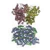
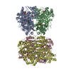
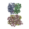
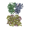
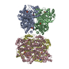
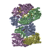
- Assembly
| Deposited unit | 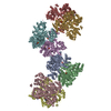
| ||||||||
|---|---|---|---|---|---|---|---|---|---|
| 1 | 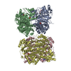
| ||||||||
| 2 | 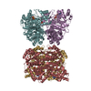
| ||||||||
| Unit cell |
| ||||||||
| Details | TETRAMER BY GEL FILTRATION AND EM DATA |
-Components
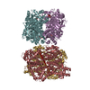
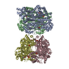
-Protein , 1 types, 8 molecules ABCDEFGH
| #1: Protein | Mass: 89144.727 Da / Num. of mol.: 8 Source method: isolated from a genetically manipulated source Details: platelet / Source: (gene. exp.) Homo sapiens (human) / Gene: PFKP, PFKF / Details (production host): Bac-to-Bac Expression system / Production host:  unidentified baculovirus / References: UniProt: Q01813, 6-phosphofructokinase unidentified baculovirus / References: UniProt: Q01813, 6-phosphofructokinase |
|---|
-Non-polymers , 5 types, 182 molecules 


| #2: Chemical | ChemComp-ATP /  Mass: 507.181 Da / Num. of mol.: 8 / Source method: obtained synthetically / Formula: C10H16N5O13P3 / Comment: ATP, energy-carrying molecule*YM Mass: 507.181 Da / Num. of mol.: 8 / Source method: obtained synthetically / Formula: C10H16N5O13P3 / Comment: ATP, energy-carrying molecule*YM#3: Chemical | ChemComp-MG /  Mass: 24.305 Da / Num. of mol.: 15 / Source method: obtained synthetically / Formula: Mg Mass: 24.305 Da / Num. of mol.: 15 / Source method: obtained synthetically / Formula: Mg#4: Chemical | ChemComp-PO4 /  Mass: 94.971 Da / Num. of mol.: 12 / Source method: obtained synthetically / Formula: PO4 Mass: 94.971 Da / Num. of mol.: 12 / Source method: obtained synthetically / Formula: PO4#5: Chemical |  Mass: 58.933 Da / Num. of mol.: 2 / Source method: obtained synthetically / Formula: Co Mass: 58.933 Da / Num. of mol.: 2 / Source method: obtained synthetically / Formula: Co#6: Water | ChemComp-HOH / | Mass: 18.015 Da / Num. of mol.: 145 / Source method: isolated from a natural source / Formula: H2O |
|---|
-Experimental details
-Experiment
| Experiment | Method: X-RAY DIFFRACTION |
|---|
- Sample preparation
| Crystal | Density Matthews: 2.68 Å3/Da / Density % sol: 54.07 % |
|---|---|
| Crystal grow | Temperature: 293 K / Method: microbatch / pH: 7 Details: 200 mM potassium thiocyanate, pH 7, and 20% (w/v) PEG 3350 |
-Data collection
| Diffraction | Mean temperature: 100 K |
|---|---|
| Diffraction source | Source: SYNCHROTRON / Site: NSLS  / Beamline: X4C / Wavelength: 0.97907 Å / Beamline: X4C / Wavelength: 0.97907 Å |
| Detector | Type: MAR CCD 165 mm / Detector: CCD / Date: Sep 17, 2013 |
| Radiation | Monochromator: Si / Protocol: SINGLE WAVELENGTH / Monochromatic (M) / Laue (L): M / Scattering type: x-ray |
| Radiation wavelength | Wavelength: 0.97907 Å / Relative weight: 1 |
| Reflection | Resolution: 3.1→49.69 Å / Num. obs: 121790 / % possible obs: 93 % / Redundancy: 6.4 % / Biso Wilson estimate: 53.7 Å2 / Rsym value: 0.084 / Net I/σ(I): 13.9 |
| Reflection shell | Resolution: 3.1→3.21 Å / Redundancy: 5.3 % / Rmerge(I) obs: 0.684 / Mean I/σ(I) obs: 1.84 / % possible all: 84.1 |
- Processing
| Software |
| ||||||||||||||||||||||||||||||||||||||||||||||||||||||||||||
|---|---|---|---|---|---|---|---|---|---|---|---|---|---|---|---|---|---|---|---|---|---|---|---|---|---|---|---|---|---|---|---|---|---|---|---|---|---|---|---|---|---|---|---|---|---|---|---|---|---|---|---|---|---|---|---|---|---|---|---|---|---|
| Refinement | Method to determine structure: MOLECULAR REPLACEMENT Starting model: 3O8L Resolution: 3.1→49.69 Å / Rfactor Rfree error: 0.002 / Data cutoff high absF: 451885.17 / Data cutoff low absF: 0 / Isotropic thermal model: RESTRAINED / Cross valid method: THROUGHOUT / σ(F): 0
| ||||||||||||||||||||||||||||||||||||||||||||||||||||||||||||
| Solvent computation | Solvent model: FLAT MODEL / Bsol: 16.4025 Å2 / ksol: 0.3 e/Å3 | ||||||||||||||||||||||||||||||||||||||||||||||||||||||||||||
| Displacement parameters | Biso mean: 62.8 Å2
| ||||||||||||||||||||||||||||||||||||||||||||||||||||||||||||
| Refine analyze |
| ||||||||||||||||||||||||||||||||||||||||||||||||||||||||||||
| Refinement step | Cycle: LAST / Resolution: 3.1→49.69 Å /
| ||||||||||||||||||||||||||||||||||||||||||||||||||||||||||||
| Refine LS restraints |
| ||||||||||||||||||||||||||||||||||||||||||||||||||||||||||||
| LS refinement shell | Resolution: 3.1→3.29 Å / Rfactor Rfree error: 0.01 / Total num. of bins used: 6
|
