 Movie
Movie Controller

[English] 日本語
 Yorodumi
- PDB-4wl0: Ligand-free structure of human platelet phosphofructokinase in an... -
+ Open data

- Basic information
| Entry | Database: PDB / ID: 4wl0 | ||||||
|---|---|---|---|---|---|---|---|
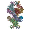
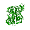
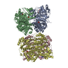
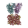
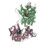
| Title | Ligand-free structure of human platelet phosphofructokinase in an R-state, crystal form I | ||||||
 Components Components | ATP-dependent 6-phosphofructokinase, platelet type | ||||||
 Keywords Keywords | TRANSFERASE / human platelet phosphofructokinase / fructose 6-phosphate / main regulator of glycolysis | ||||||
| Function / homology |  Function and homology information Function and homology information6-phosphofructokinase complex / 6-phosphofructokinase / 6-phosphofructokinase activity / fructose-6-phosphate binding / fructose 1,6-bisphosphate metabolic process / fructose 6-phosphate metabolic process / Glycolysis / canonical glycolysis / cellular response to leukemia inhibitory factor / cadherin binding ...6-phosphofructokinase complex / 6-phosphofructokinase / 6-phosphofructokinase activity / fructose-6-phosphate binding / fructose 1,6-bisphosphate metabolic process / fructose 6-phosphate metabolic process / Glycolysis / canonical glycolysis / cellular response to leukemia inhibitory factor / cadherin binding / protein-containing complex binding / extracellular exosome / ATP binding / membrane / metal ion binding / identical protein binding / nucleus / cytoplasm / cytosol Similarity search - Function | ||||||
| Biological species |  Homo sapiens (human) Homo sapiens (human) | ||||||
| Method |  X-RAY DIFFRACTION / SYNCHROTRON / MOLECULAR REPLACEMENT / molecular replacement / Resolution: 2.89 Å X-RAY DIFFRACTION / SYNCHROTRON / MOLECULAR REPLACEMENT / molecular replacement / Resolution: 2.89 Å | ||||||
| Model details | N- and C-terminal truncation (N=25, C=22 aa), N-terminal Strep-tag and EK-site | ||||||
 Authors Authors | Kloos, M. / Strater, N. | ||||||
| Funding support |  Germany, 1items Germany, 1items
| ||||||
 Citation Citation | Journal: Biochem.J. / Year: 2015 Title: Crystal structure of human platelet phosphofructokinase-1 locked in an activated conformation. Authors: Kloos, M. / Bruser, A. / Kirchberger, J. / Schoneberg, T. / Strater, N. #1: Journal: Acta Crystallogr F Struct Biol Commun / Year: 2014Title: Crystallization and preliminary crystallographic analysis of human muscle phosphofructokinase, the main regulator of glycolysis. Authors: Kloos, M. / Brueser, A. / Kirchberger, J. / Schoeneberg, T. / Straeter, N. #2: Journal: FASEB J. / Year: 2011Title: Molecular architecture and structural basis of allosteric regulation of eukaryotic phosphofructokinases. Authors: Straeter, N. / Marek, S. / Kuettner, E.B. / Kloos, M. / Keim, A. / Brueser, A. / Kirchberger, J. / Schoeneberg, T. | ||||||
| History |
|
- Structure visualization
| Structure viewer | Molecule: MolmilJmol/JSmol |
|---|
- Downloads & links
-Download
| PDBx/mmCIF format | 4wl0.cif.gz | 1.1 MB | Display | PDBx/mmCIF format |
|---|---|---|---|---|
| PDB format | pdb4wl0.ent.gz | 935.1 KB | Display | PDB format |
| PDBx/mmJSON format | 4wl0.json.gz | Tree view | PDBx/mmJSON format | |
| Others |  Other downloads Other downloads |
-Validation report
| Arichive directory | https://data.pdbj.org/pub/pdb/validation_reports/wl/4wl0ftp://data.pdbj.org/pub/pdb/validation_reports/wl/4wl0 | HTTPS FTP |
|---|
-Related structure data


-Links
 PDBj
PDBj

- Assembly
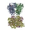
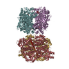
| Deposited unit | 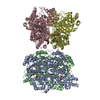
| |||||||||||||||||||||||||||||||||||
|---|---|---|---|---|---|---|---|---|---|---|---|---|---|---|---|---|---|---|---|---|---|---|---|---|---|---|---|---|---|---|---|---|---|---|---|---|
| 1 |
| |||||||||||||||||||||||||||||||||||
| Unit cell |
| |||||||||||||||||||||||||||||||||||
| Noncrystallographic symmetry (NCS) | NCS domain:
NCS domain segments: Component-ID: 1 / Ens-ID: 1 / Beg auth comp-ID: SER / Beg label comp-ID: SER / End auth comp-ID: ALA / End label comp-ID: ALA / Auth seq-ID: 25 - 762 / Label seq-ID: 24 - 761
| |||||||||||||||||||||||||||||||||||
| Details | biological unit is the same as asym. |
-Components
| #1: Protein | Mass: 83161.023 Da / Num. of mol.: 4 / Fragment: UNP residues 26-762 Source method: isolated from a genetically manipulated source Details: truncated humane platelet Pfk plasmid start: mas N-terminal Strep-tag: wshpqfek linker: ga EK-site: ddddk restriction site: vpdpts N-terminal start: aigvl N-terminal start: aa 26 C-terminal end: aa 762 Source: (gene. exp.) Homo sapiens (human) / Gene: PFKP, PFKF / Plasmid: pET51b / Production host:  #2: Chemical | ChemComp-PO4 /   Mass: 94.971 Da / Num. of mol.: 8 / Source method: obtained synthetically / Formula: PO4 Mass: 94.971 Da / Num. of mol.: 8 / Source method: obtained synthetically / Formula: PO4 |
|---|
-Experimental details
-Experiment
| Experiment | Method: X-RAY DIFFRACTION / Number of used crystals: 1 |
|---|
- Sample preparation
| Crystal | Density Matthews: 2.66 Å3/Da / Density % sol: 53.7 % |
|---|---|
| Crystal grow | Temperature: 292 K / Method: vapor diffusion, hanging drop / pH: 6 Details: 0.1 M KCl, 0.025 M MgCl2, 0.05 M Na Cacodylate, 15% iso-Propanol |
-Data collection
| Diffraction | Mean temperature: 100 K | ||||||||||||||||||||||||||||||||||||||||||||||||||||||||||||||||||||||||||||||||||||||||||||||||||||
|---|---|---|---|---|---|---|---|---|---|---|---|---|---|---|---|---|---|---|---|---|---|---|---|---|---|---|---|---|---|---|---|---|---|---|---|---|---|---|---|---|---|---|---|---|---|---|---|---|---|---|---|---|---|---|---|---|---|---|---|---|---|---|---|---|---|---|---|---|---|---|---|---|---|---|---|---|---|---|---|---|---|---|---|---|---|---|---|---|---|---|---|---|---|---|---|---|---|---|---|---|---|
| Diffraction source | Source: SYNCHROTRON / Site: BESSY / Beamline: 14.1 / Wavelength: 0.91841 Å | ||||||||||||||||||||||||||||||||||||||||||||||||||||||||||||||||||||||||||||||||||||||||||||||||||||
| Detector | Type: DECTRIS PILATUS 6M / Detector: PIXEL / Date: Oct 15, 2013 | ||||||||||||||||||||||||||||||||||||||||||||||||||||||||||||||||||||||||||||||||||||||||||||||||||||
| Radiation | Monochromator: Si-111 crysta / Protocol: SINGLE WAVELENGTH / Monochromatic (M) / Laue (L): M / Scattering type: x-ray | ||||||||||||||||||||||||||||||||||||||||||||||||||||||||||||||||||||||||||||||||||||||||||||||||||||
| Radiation wavelength | Wavelength: 0.91841 Å / Relative weight: 1 | ||||||||||||||||||||||||||||||||||||||||||||||||||||||||||||||||||||||||||||||||||||||||||||||||||||
| Reflection | Resolution: 2.89→50 Å / Num. obs: 79810 / % possible obs: 99.3 % / Observed criterion σ(I): -3 / Redundancy: 3.6 % / Biso Wilson estimate: 67.66 Å2 / Rmerge F obs: 0.998 / Rmerge(I) obs: 0.077 / Rrim(I) all: 0.091 / Χ2: 1.061 / Net I/σ(I): 13.1 / Num. measured all: 289757 | ||||||||||||||||||||||||||||||||||||||||||||||||||||||||||||||||||||||||||||||||||||||||||||||||||||
| Reflection shell | Diffraction-ID: 1 / Rejects: _
|
-Phasing
| Phasing | Method: molecular replacement | |||||||||
|---|---|---|---|---|---|---|---|---|---|---|
| Phasing MR | Model details: Phaser MODE: MR_AUTO
|
- Processing
| Software |
| ||||||||||||||||||||||||||||||||||||||||||||||||||||||||||||||||||||||||||||||||||||||||||||||||||||||||||||||||||||||||||||||||||||||||||||||||||||||||||||||||||||||||||||||||||||||||||||||||||||||||||||||||||
|---|---|---|---|---|---|---|---|---|---|---|---|---|---|---|---|---|---|---|---|---|---|---|---|---|---|---|---|---|---|---|---|---|---|---|---|---|---|---|---|---|---|---|---|---|---|---|---|---|---|---|---|---|---|---|---|---|---|---|---|---|---|---|---|---|---|---|---|---|---|---|---|---|---|---|---|---|---|---|---|---|---|---|---|---|---|---|---|---|---|---|---|---|---|---|---|---|---|---|---|---|---|---|---|---|---|---|---|---|---|---|---|---|---|---|---|---|---|---|---|---|---|---|---|---|---|---|---|---|---|---|---|---|---|---|---|---|---|---|---|---|---|---|---|---|---|---|---|---|---|---|---|---|---|---|---|---|---|---|---|---|---|---|---|---|---|---|---|---|---|---|---|---|---|---|---|---|---|---|---|---|---|---|---|---|---|---|---|---|---|---|---|---|---|---|---|---|---|---|---|---|---|---|---|---|---|---|---|---|---|---|---|
| Refinement | Method to determine structure: MOLECULAR REPLACEMENT Starting model: unpublished low resolution structure Resolution: 2.89→46.97 Å / FOM work R set: 0.7776 / SU ML: 0.38 / Cross valid method: FREE R-VALUE / σ(F): 1.36 / Phase error: 28.84 / Stereochemistry target values: ML
| ||||||||||||||||||||||||||||||||||||||||||||||||||||||||||||||||||||||||||||||||||||||||||||||||||||||||||||||||||||||||||||||||||||||||||||||||||||||||||||||||||||||||||||||||||||||||||||||||||||||||||||||||||
| Solvent computation | Shrinkage radii: 0.9 Å / VDW probe radii: 1.11 Å / Solvent model: FLAT BULK SOLVENT MODEL | ||||||||||||||||||||||||||||||||||||||||||||||||||||||||||||||||||||||||||||||||||||||||||||||||||||||||||||||||||||||||||||||||||||||||||||||||||||||||||||||||||||||||||||||||||||||||||||||||||||||||||||||||||
| Displacement parameters | Biso max: 312.71 Å2 / Biso mean: 96.17 Å2 / Biso min: 39.06 Å2 | ||||||||||||||||||||||||||||||||||||||||||||||||||||||||||||||||||||||||||||||||||||||||||||||||||||||||||||||||||||||||||||||||||||||||||||||||||||||||||||||||||||||||||||||||||||||||||||||||||||||||||||||||||
| Refinement step | Cycle: final / Resolution: 2.89→46.97 Å
| ||||||||||||||||||||||||||||||||||||||||||||||||||||||||||||||||||||||||||||||||||||||||||||||||||||||||||||||||||||||||||||||||||||||||||||||||||||||||||||||||||||||||||||||||||||||||||||||||||||||||||||||||||
| Refine LS restraints |
| ||||||||||||||||||||||||||||||||||||||||||||||||||||||||||||||||||||||||||||||||||||||||||||||||||||||||||||||||||||||||||||||||||||||||||||||||||||||||||||||||||||||||||||||||||||||||||||||||||||||||||||||||||
| Refine LS restraints NCS |
| ||||||||||||||||||||||||||||||||||||||||||||||||||||||||||||||||||||||||||||||||||||||||||||||||||||||||||||||||||||||||||||||||||||||||||||||||||||||||||||||||||||||||||||||||||||||||||||||||||||||||||||||||||
| LS refinement shell | Refine-ID: X-RAY DIFFRACTION / Total num. of bins used: 29
| ||||||||||||||||||||||||||||||||||||||||||||||||||||||||||||||||||||||||||||||||||||||||||||||||||||||||||||||||||||||||||||||||||||||||||||||||||||||||||||||||||||||||||||||||||||||||||||||||||||||||||||||||||
| Refinement TLS params. | Method: refined / Refine-ID: X-RAY DIFFRACTION
| ||||||||||||||||||||||||||||||||||||||||||||||||||||||||||||||||||||||||||||||||||||||||||||||||||||||||||||||||||||||||||||||||||||||||||||||||||||||||||||||||||||||||||||||||||||||||||||||||||||||||||||||||||
| Refinement TLS group |
|
