 Movie
Movie Controller


+ Open data

- Basic information
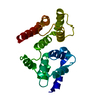
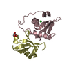
| Entry | Database: PDB / ID: 7lvj | ||||||
|---|---|---|---|---|---|---|---|
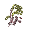
| Title | CASP8 isoform G DED domain | ||||||
 Components Components | Isoform 9 of Caspase-8 | ||||||
 Keywords Keywords | HYDROLASE / CASP8g / CASP8 / GENE ID: 841 / CASP8G DED | ||||||
| Function / homology | caspase-8 / Isoform 9 of Caspase-8 Function and homology information Function and homology information | ||||||
| Biological species |  Homo sapiens (human) Homo sapiens (human) | ||||||
| Method |  X-RAY DIFFRACTION / SYNCHROTRON / MOLECULAR REPLACEMENT / Resolution: 1.5 Å X-RAY DIFFRACTION / SYNCHROTRON / MOLECULAR REPLACEMENT / Resolution: 1.5 Å | ||||||
 Authors Authors | Weichert, K. / Lu, F. / Kodandapani, L. / Sauder, J.M. | ||||||
 Citation Citation | Journal: ACR Open Rheumatol / Year: 2022 Title: Caspase-8 Variant G Regulates Rheumatoid Arthritis Fibroblast-Like Synoviocyte Aggressive Behavior. Authors: Ansalone, C. / Ainsworth, R.I. / Nygaard, G. / Ai, R. / Prideaux, E.B. / Hammaker, D. / Perumal, N.B. / Weichert, K. / Tung, F. / Kodandapani, L. / Sauder, J.M. / Mertsching, E.C. / ...Authors: Ansalone, C. / Ainsworth, R.I. / Nygaard, G. / Ai, R. / Prideaux, E.B. / Hammaker, D. / Perumal, N.B. / Weichert, K. / Tung, F. / Kodandapani, L. / Sauder, J.M. / Mertsching, E.C. / Benschop, R.J. / Boyle, D.L. / Wang, W. / Firestein, G.S. | ||||||
| History |
|
- Structure visualization
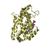
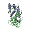
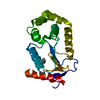
| Structure viewer | Molecule: MolmilJmol/JSmol |
|---|
- Downloads & links
-Download
| PDBx/mmCIF format | 7lvj.cif.gz | 58.8 KB | Display | PDBx/mmCIF format |
|---|---|---|---|---|
| PDB format | pdb7lvj.ent.gz | 41.2 KB | Display | PDB format |
| PDBx/mmJSON format | 7lvj.json.gz | Tree view | PDBx/mmJSON format | |
| Others |  Other downloads Other downloads |
-Validation report
| Arichive directory | https://data.pdbj.org/pub/pdb/validation_reports/lv/7lvjftp://data.pdbj.org/pub/pdb/validation_reports/lv/7lvj | HTTPS FTP |
|---|
-Related structure data


-Links
 PDBj
PDBj- Assembly
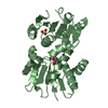
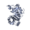
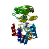
| Deposited unit | 
| ||||||||
|---|---|---|---|---|---|---|---|---|---|
| 1 |
| ||||||||
| Unit cell |
| ||||||||
| Components on special symmetry positions |
|
-Components
| #1: Protein | Mass: 30014.520 Da / Num. of mol.: 1 Source method: isolated from a genetically manipulated source Source: (gene. exp.) Homo sapiens (human) / Gene: CASP8, MCH5 / Variant: Ge. / Production host:  |
|---|---|
| #2: Water | ChemComp-HOH /  Mass: 18.015 Da / Num. of mol.: 187 / Source method: isolated from a natural source / Formula: H2O Mass: 18.015 Da / Num. of mol.: 187 / Source method: isolated from a natural source / Formula: H2O |
-Experimental details
-Experiment
| Experiment | Method: X-RAY DIFFRACTION / Number of used crystals: 1 |
|---|
- Sample preparation
| Crystal | Density Matthews: 1.96 Å3/Da / Density % sol: 37.22 % |
|---|---|
| Crystal grow | Temperature: 281 K / Method: vapor diffusion, sitting drop / pH: 8 Details: 20% PEG 3350, 200mM Potassium Sodium Tartrate tetrahydrate Temp details: 8C |
-Data collection
| Diffraction | Mean temperature: 100 K / Serial crystal experiment: N | ||||||||||||||||||||||||||||||
|---|---|---|---|---|---|---|---|---|---|---|---|---|---|---|---|---|---|---|---|---|---|---|---|---|---|---|---|---|---|---|---|
| Diffraction source | Source: SYNCHROTRON / Site: ESRF  / Beamline: ID30B / Wavelength: 0.984 Å / Beamline: ID30B / Wavelength: 0.984 Å | ||||||||||||||||||||||||||||||
| Detector | Type: DECTRIS PILATUS 6M / Detector: PIXEL / Date: Sep 7, 2020 | ||||||||||||||||||||||||||||||
| Radiation | Protocol: SINGLE WAVELENGTH / Monochromatic (M) / Laue (L): M / Scattering type: x-ray | ||||||||||||||||||||||||||||||
| Radiation wavelength | Wavelength: 0.984 Å / Relative weight: 1 | ||||||||||||||||||||||||||||||
| Reflection | Resolution: 1.5→35.98 Å / Num. obs: 36268 / % possible obs: 97.3 % / Redundancy: 2.8 % / CC1/2: 0.996 / Rmerge(I) obs: 0.054 / Rpim(I) all: 0.037 / Rrim(I) all: 0.066 / Net I/σ(I): 10.6 / Num. measured all: 101116 / Scaling rejects: 1 | ||||||||||||||||||||||||||||||
| Reflection shell | Diffraction-ID: 1
|
- Processing
| Software |
| |||||||||||||||||||||||||||||||||||||||||||||
|---|---|---|---|---|---|---|---|---|---|---|---|---|---|---|---|---|---|---|---|---|---|---|---|---|---|---|---|---|---|---|---|---|---|---|---|---|---|---|---|---|---|---|---|---|---|---|
| Refinement | Method to determine structure: MOLECULAR REPLACEMENT Starting model: Internal structure Resolution: 1.5→30 Å / Cor.coef. Fo:Fc: 0.961 / Cor.coef. Fo:Fc free: 0.949 / SU B: 1.485 / SU ML: 0.056 / Cross valid method: THROUGHOUT / σ(F): 0 / ESU R: 0.078 / ESU R Free: 0.081 / Stereochemistry target values: MAXIMUM LIKELIHOOD Details: HYDROGENS HAVE BEEN USED IF PRESENT IN THE INPUT U VALUES : REFINED INDIVIDUALLY
| |||||||||||||||||||||||||||||||||||||||||||||
| Solvent computation | Ion probe radii: 0.8 Å / Shrinkage radii: 0.8 Å / VDW probe radii: 1.2 Å / Solvent model: MASK | |||||||||||||||||||||||||||||||||||||||||||||
| Displacement parameters | Biso max: 90.33 Å2 / Biso mean: 23.198 Å2 / Biso min: 10.38 Å2
| |||||||||||||||||||||||||||||||||||||||||||||
| Refinement step | Cycle: final / Resolution: 1.5→30 Å
| |||||||||||||||||||||||||||||||||||||||||||||
| Refine LS restraints |
| |||||||||||||||||||||||||||||||||||||||||||||
| LS refinement shell | Resolution: 1.5→1.539 Å / Rfactor Rfree error: 0 / Total num. of bins used: 20
|
