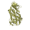
 Movie
Movie Controller


+ Open data

- Basic information
| Entry | Database: PDB / ID: 7lsa | ||||||
|---|---|---|---|---|---|---|---|
| Title | Ruminococcus bromii Amy12 with maltoheptaose | ||||||
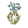
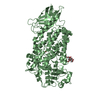
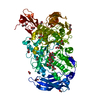
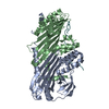
 Components Components | Pullulanase | ||||||
 Keywords Keywords | HYDROLASE / GH13 / pullulanase / CBM48 / complex | ||||||
| Function / homology |  Function and homology information Function and homology informationpullulanase / pullulanase activity / cellulose catabolic process / extracellular region / metal ion binding Similarity search - Function | ||||||
| Biological species |  Ruminococcus bromii (bacteria) Ruminococcus bromii (bacteria) | ||||||
| Method |  X-RAY DIFFRACTION / SYNCHROTRON / MOLECULAR REPLACEMENT / molecular replacement / Resolution: 1.76 Å X-RAY DIFFRACTION / SYNCHROTRON / MOLECULAR REPLACEMENT / molecular replacement / Resolution: 1.76 Å | ||||||
 Authors Authors | Koropatkin, N.M. / Cockburn, D.W. / Brown, H.A. / Kibler, R.D. | ||||||
| Funding support |  United States, 1items United States, 1items
| ||||||
 Citation Citation | Journal: J.Struct.Biol. / Year: 2021 Title: Structure and substrate recognition by the Ruminococcus bromii amylosome pullulanases. Authors: Cockburn, D.W. / Kibler, R. / Brown, H.A. / Duvall, R. / Morais, S. / Bayer, E. / Koropatkin, N.M. | ||||||
| History |
|
- Structure visualization
| Structure viewer | Molecule: MolmilJmol/JSmol |
|---|
- Downloads & links
-Download
| PDBx/mmCIF format | 7lsa.cif.gz | 339.6 KB | Display | PDBx/mmCIF format |
|---|---|---|---|---|
| PDB format | pdb7lsa.ent.gz | 274.4 KB | Display | PDB format |
| PDBx/mmJSON format | 7lsa.json.gz | Tree view | PDBx/mmJSON format | |
| Others |  Other downloads Other downloads |
-Validation report
| Arichive directory | https://data.pdbj.org/pub/pdb/validation_reports/ls/7lsaftp://data.pdbj.org/pub/pdb/validation_reports/ls/7lsa | HTTPS FTP |
|---|
-Related structure data
| Related structure data | 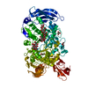 7lsrC 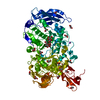 7lstC  7lsuC  2wanS S: Starting model for refinement C: citing same article ( |
|---|---|
| Similar structure data |


-Links
 PDBj
PDBj


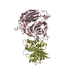
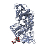
- Assembly
| Deposited unit | 
| ||||||||
|---|---|---|---|---|---|---|---|---|---|
| 1 | 
| ||||||||
| 2 | 
| ||||||||
| Unit cell |
|
-Components
-Protein , 1 types, 1 molecules A
| #1: Protein | Mass: 87172.875 Da / Num. of mol.: 1 Source method: isolated from a genetically manipulated source Source: (gene. exp.) Ruminococcus bromii (bacteria) / Gene: pulA_2, RBL236_00821 / Production host: |
|---|
-Sugars , 2 types, 2 molecules
| #2: Polysaccharide | alpha-D-glucopyranose-(1-4)-alpha-D-glucopyranose-(1-4)-alpha-D-glucopyranose-(1-4)-alpha-D- ...alpha-D-glucopyranose-(1-4)-alpha-D-glucopyranose-(1-4)-alpha-D-glucopyranose-(1-4)-alpha-D-glucopyranose-(1-4)-alpha-D-glucopyranose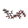  Source method: isolated from a genetically manipulated source Details: oligosaccharide / References: alpha-maltopentaose |
|---|---|
| #3: Polysaccharide | alpha-D-glucopyranose-(1-4)-alpha-D-glucopyranose-(1-4)-alpha-D-glucopyranose  Source method: isolated from a genetically manipulated source Details: oligosaccharide / References: alpha-maltotriose |
-Non-polymers , 4 types, 730 molecules 


| #4: Chemical | ChemComp-GOL /  Mass: 92.094 Da / Num. of mol.: 8 / Source method: obtained synthetically / Formula: C3H8O3 Mass: 92.094 Da / Num. of mol.: 8 / Source method: obtained synthetically / Formula: C3H8O3#5: Chemical | ChemComp-PEG / |  Mass: 106.120 Da / Num. of mol.: 1 / Source method: obtained synthetically / Formula: C4H10O3 Mass: 106.120 Da / Num. of mol.: 1 / Source method: obtained synthetically / Formula: C4H10O3#6: Chemical | ChemComp-CA / |  Mass: 40.078 Da / Num. of mol.: 1 / Source method: obtained synthetically / Formula: Ca Mass: 40.078 Da / Num. of mol.: 1 / Source method: obtained synthetically / Formula: Ca#7: Water | ChemComp-HOH / | Mass: 18.015 Da / Num. of mol.: 720 / Source method: isolated from a natural source / Formula: H2O |
|---|
-Details
| Has ligand of interest | Y |
|---|
-Experimental details
-Experiment
| Experiment | Method: X-RAY DIFFRACTION / Number of used crystals: 1 |
|---|
- Sample preparation
| Crystal | Density Matthews: 2.21 Å3/Da / Density % sol: 44.22 % |
|---|---|
| Crystal grow | Temperature: 277 K / Method: vapor diffusion, hanging drop / pH: 6.5 Details: well solution: 16% PEG 3350, 4% glycerol, 0.3 ammonium acetate, and 0.1 M Bis-Tris pH 6.5; protein: 8.75 - 8.9 mg/ml Amy12 with 10mM maltoheptaose Temp details: cold room |
-Data collection
| Diffraction | Mean temperature: 120 K / Ambient temp details: cryogenic / Serial crystal experiment: N |
|---|---|
| Diffraction source | Source: SYNCHROTRON / Site: APS / Beamline: 21-ID-F / Wavelength: 0.979 Å |
| Detector | Type: RAYONIX MX300-HS / Detector: CCD / Date: Feb 11, 2015 |
| Radiation | Monochromator: C(111) / Protocol: SINGLE WAVELENGTH / Monochromatic (M) / Laue (L): M / Scattering type: x-ray |
| Radiation wavelength | Wavelength: 0.979 Å / Relative weight: 1 |
| Reflection | Resolution: 1.76→48.46 Å / Num. obs: 74259 / % possible obs: 95.9 % / Redundancy: 4.7 % / CC1/2: 0.99 / Net I/σ(I): 8.6 |
| Reflection shell | Resolution: 1.76→1.82 Å / Num. unique obs: 5901 / CC1/2: 0.26 |
-Phasing
| Phasing | Method: molecular replacement |
|---|
- Processing
| Software |
| |||||||||||||||||||||||||||||||||||||||||||||||||||||||||||||||||||||||||||
|---|---|---|---|---|---|---|---|---|---|---|---|---|---|---|---|---|---|---|---|---|---|---|---|---|---|---|---|---|---|---|---|---|---|---|---|---|---|---|---|---|---|---|---|---|---|---|---|---|---|---|---|---|---|---|---|---|---|---|---|---|---|---|---|---|---|---|---|---|---|---|---|---|---|---|---|---|
| Refinement | Method to determine structure: MOLECULAR REPLACEMENT Starting model: 2WAN Resolution: 1.76→48.46 Å / Cor.coef. Fo:Fc: 0.969 / Cor.coef. Fo:Fc free: 0.947 / SU B: 7.334 / SU ML: 0.093 / Cross valid method: THROUGHOUT / σ(F): 0 / ESU R: 0.222 / ESU R Free: 0.114 / Stereochemistry target values: MAXIMUM LIKELIHOOD Details: HYDROGENS HAVE BEEN ADDED IN THE RIDING POSITIONS U VALUES : WITH TLS ADDED
| |||||||||||||||||||||||||||||||||||||||||||||||||||||||||||||||||||||||||||
| Solvent computation | Ion probe radii: 0.8 Å / Shrinkage radii: 0.8 Å / VDW probe radii: 1.2 Å / Solvent model: MASK | |||||||||||||||||||||||||||||||||||||||||||||||||||||||||||||||||||||||||||
| Displacement parameters | Biso max: 53.86 Å2 / Biso mean: 19.913 Å2 / Biso min: 10.59 Å2
| |||||||||||||||||||||||||||||||||||||||||||||||||||||||||||||||||||||||||||
| Refinement step | Cycle: final / Resolution: 1.76→48.46 Å
| |||||||||||||||||||||||||||||||||||||||||||||||||||||||||||||||||||||||||||
| Refine LS restraints |
| |||||||||||||||||||||||||||||||||||||||||||||||||||||||||||||||||||||||||||
| LS refinement shell | Resolution: 1.76→1.806 Å / Rfactor Rfree error: 0 / Total num. of bins used: 20
| |||||||||||||||||||||||||||||||||||||||||||||||||||||||||||||||||||||||||||
| Refinement TLS params. | Method: refined / Origin x: 62.4534 Å / Origin y: 99.9484 Å / Origin z: 188.2997 Å
|
