 Movie
Movie Controller


+ Open data

- Basic information
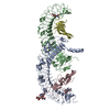
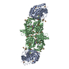
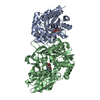
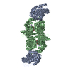
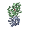
| Entry | Database: PDB / ID: 2vdu | ||||||
|---|---|---|---|---|---|---|---|
| Title | Structure of trm8-trm82, THE YEAST TRNA m7G methylation complex | ||||||
 Components Components |
| ||||||
 Keywords Keywords | TRANSFERASE / S-ADENOSYL-L-METHIONINE / TRNA PROCESSING / PHOSPHORYLATION / METHYLTRANSFERASE / M7G / TRNA / SPOUT MT / WD REPEAT | ||||||
| Function / homology |  Function and homology information Function and homology informationtRNA (m7G46) methyltransferase complex / tRNA (guanine-N7)-methylation / RNA (guanine-N7)-methylation / tRNA (guanine46-N7)-methyltransferase / tRNA (guanine(46)-N7)-methyltransferase activity / tRNA methyltransferase complex / tRNA modification / tRNA methylation / enzyme activator activity / tRNA binding ...tRNA (m7G46) methyltransferase complex / tRNA (guanine-N7)-methylation / RNA (guanine-N7)-methylation / tRNA (guanine46-N7)-methyltransferase / tRNA (guanine(46)-N7)-methyltransferase activity / tRNA methyltransferase complex / tRNA modification / tRNA methylation / enzyme activator activity / tRNA binding / nucleoplasm / nucleus / cytosol Similarity search - Function | ||||||
| Biological species |  | ||||||
| Method |  X-RAY DIFFRACTION / SYNCHROTRON / SAD / Resolution: 2.4 Å X-RAY DIFFRACTION / SYNCHROTRON / SAD / Resolution: 2.4 Å | ||||||
 Authors Authors | Leulliot, N. / Chaillet, M. / Durand, D. / Ulryck, N. / Blondeau, K. / Van Tilbeurgh, H. | ||||||
 Citation Citation | Journal: Structure / Year: 2008 Title: Structure of the Yeast tRNA M7G Methylation Complex. Authors: Leulliot, N. / Chaillet, M. / Durand, D. / Ulryck, N. / Blondeau, K. / Van Tilbeurgh, H. | ||||||
| History |
|
- Structure visualization
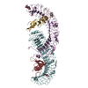
| Structure viewer | Molecule: MolmilJmol/JSmol |
|---|
- Downloads & links
-Download
| PDBx/mmCIF format | 2vdu.cif.gz | 246.1 KB | Display | PDBx/mmCIF format |
|---|---|---|---|---|
| PDB format | pdb2vdu.ent.gz | 197.7 KB | Display | PDB format |
| PDBx/mmJSON format | 2vdu.json.gz | Tree view | PDBx/mmJSON format | |
| Others |  Other downloads Other downloads |
-Validation report
| Arichive directory | https://data.pdbj.org/pub/pdb/validation_reports/vd/2vduftp://data.pdbj.org/pub/pdb/validation_reports/vd/2vdu | HTTPS FTP |
|---|
-Related structure data


-Links
 PDBj
PDBj

- Assembly
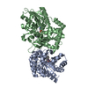
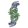
| Deposited unit | 
| ||||||||
|---|---|---|---|---|---|---|---|---|---|
| 1 |
| ||||||||
| 2 | 
| ||||||||
| 3 | 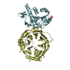
| ||||||||
| Unit cell |
|
-Components
| #1: Protein | Mass: 51383.844 Da / Num. of mol.: 2 Source method: isolated from a genetically manipulated source Source: (gene. exp.) Production host:  #2: Protein | Mass: 29796.221 Da / Num. of mol.: 2 / Fragment: 46-END, RESIDUES 39-286 Source method: isolated from a genetically manipulated source Details: 46 N-TERMNIAL TRUNCATION Source: (gene. exp.) Production host: References: UniProt: Q12009, tRNA (guanine46-N7)-methyltransferase #3: Chemical |   Mass: 94.971 Da / Num. of mol.: 2 / Source method: obtained synthetically / Formula: PO4 Mass: 94.971 Da / Num. of mol.: 2 / Source method: obtained synthetically / Formula: PO4#4: Water | ChemComp-HOH / |  Mass: 18.015 Da / Num. of mol.: 259 / Source method: isolated from a natural source / Formula: H2O Mass: 18.015 Da / Num. of mol.: 259 / Source method: isolated from a natural source / Formula: H2OSequence details | N-TERMINAL 46 RESIDUE TRUNCATION | |
|---|
-Experimental details
-Experiment
| Experiment | Method: X-RAY DIFFRACTION |
|---|
- Sample preparation
| Crystal | Density Matthews: 2.74 Å3/Da / Density % sol: 45.56 % / Description: NONE |
|---|
-Data collection
| Diffraction | Mean temperature: 100 K |
|---|---|
| Diffraction source | Source: SYNCHROTRON / Site: ESRF  / Beamline: ID14-4 / Wavelength: 0.97953 / Beamline: ID14-4 / Wavelength: 0.97953 |
| Detector | Type: ADSC CCD / Detector: CCD |
| Radiation | Protocol: SINGLE WAVELENGTH / Monochromatic (M) / Laue (L): M / Scattering type: x-ray |
| Radiation wavelength | Wavelength: 0.97953 Å / Relative weight: 1 |
| Reflection | Resolution: 2.4→20 Å / Num. obs: 26954 / % possible obs: 98.2 % / Observed criterion σ(I): 1.5 / Redundancy: 3.7 % / Rmerge(I) obs: 0.08 / Net I/σ(I): 12.2 |
| Reflection shell | Resolution: 2.3→2.4 Å / Redundancy: 3.7 % / Rmerge(I) obs: 0.69 / Mean I/σ(I) obs: 1.5 / % possible all: 99.1 |
- Processing
| Software |
| ||||||||||||||||
|---|---|---|---|---|---|---|---|---|---|---|---|---|---|---|---|---|---|
| Refinement | Method to determine structure: SAD Starting model: NONE Resolution: 2.4→20 Å / Stereochemistry target values: ML
| ||||||||||||||||
| Refinement step | Cycle: LAST / Resolution: 2.4→20 Å
|
