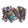
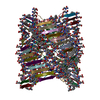
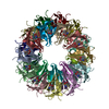
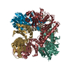
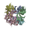
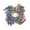
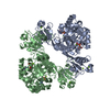
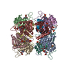
Journal: Matter / Year: 2021 Title: Deterministic chaos in the self-assembly of β sheet nanotubes from an amphipathic oligopeptide. Authors: Fengbin Wang / Ordy Gnewou / Shengyuan Wang / Tomasz Osinski / Xiaobing Zuo / Edward H Egelman / Vincent P Conticello / Abstract: The self-assembly of designed peptides into filaments and other higher-order structures has been the focus of intense interest because of the potential for creating new biomaterials and biomedical ...The self-assembly of designed peptides into filaments and other higher-order structures has been the focus of intense interest because of the potential for creating new biomaterials and biomedical devices. These peptide assemblies have also been used as models for understanding biological processes, such as the pathological formation of amyloid. We investigate the assembly of an octapeptide sequence, Ac-FKFEFKFE-NH, motivated by prior studies that demonstrated that this amphipathic β strand peptide self-assembled into fibrils and biocompatible hydrogels. Using high-resolution cryoelectron microscopy (cryo-EM), we are able to determine the atomic structure for two different coexisting forms of the fibrils, containing four and five β sandwich protofilaments, respectively. Surprisingly, the inner walls in both forms are parallel β sheets, while the outer walls are antiparallel β sheets. Our results demonstrate the chaotic nature of peptide self-assembly and illustrate the importance of cryo-EM structural analysis to understand the complex phase behavior of these materials at near-atomic resolution.
History
Deposition
Feb 13, 2021
Deposition site: RCSB / Processing site: RCSB
Revision 1.0
Jun 30, 2021
Provider: repository / Type: Initial release
Revision 1.0
Jun 30, 2021
Data content type: EM metadata / Data content type: EM metadata / Provider: repository / Type: Initial release
Revision 1.0
Jun 30, 2021
Data content type: Image / Data content type: Image / Provider: repository / Type: Initial release
Revision 1.0
Jun 30, 2021
Data content type: Primary map / Data content type: Primary map / Provider: repository / Type: Initial release
Revision 1.0
Jun 30, 2021
Data content type: Image / Data content type: Image / Provider: repository / Type: Initial release
Revision 1.0
Jun 30, 2021
Data content type: Primary map / Data content type: Primary map / Provider: repository / Type: Initial release
Revision 1.0
Jun 30, 2021
Data content type: Image / Data content type: Image / Provider: repository / Type: Initial release
Revision 1.0
Jun 30, 2021
Data content type: Primary map / Data content type: Primary map / Provider: repository / Type: Initial release
Revision 1.0
Jun 30, 2021
Data content type: Image / Data content type: Image / Provider: repository / Type: Initial release
Revision 1.0
Jun 30, 2021
Data content type: Primary map / Data content type: Primary map / Provider: repository / Type: Initial release
Data content type: EM metadata / Data content type: EM metadata / EM metadata / Group: Data processing / Experimental summary / Data content type: EM metadata / EM metadata / Category: em_admin / em_software / Data content type: EM metadata / EM metadata / Item: _em_admin.last_update / _em_software.name
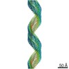
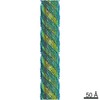
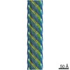
Evidence: microscopy, helical filament was observed by negative staining and Cryo-EM
Type
Name
Symmetry operation
Number
identity operation
1_555
1
Symmetry
Helical symmetry: (Circular symmetry: 2 / Dyad axis: no / N subunits divisor: 1 / Num. of operations: 144 / Rise per n subunits: 3.97 Å / Rotation per n subunits: 172.1 °)
-
Components
#1: Protein/peptide
... KFE8peptide
Mass: 1164.350 Da / Num. of mol.: 144 / Source method: obtained synthetically / Source: (synth.) synthetic construct (others)
Has ligand of interest
Y
Has protein modification
Y
-
Experimental details
-
Experiment
Experiment
Method: ELECTRON MICROSCOPY
EM experiment
Aggregation state: FILAMENT / 3D reconstruction method: helical reconstruction
In the structure databanks used in Yorodumi, some data are registered as the other names, "COVID-19 virus" and "2019-nCoV". Here are the details of the virus and the list of structure data.
Jan 31, 2019. EMDB accession codes are about to change! (news from PDBe EMDB page)
EMDB accession codes are about to change! (news from PDBe EMDB page)
The allocation of 4 digits for EMDB accession codes will soon come to an end. Whilst these codes will remain in use, new EMDB accession codes will include an additional digit and will expand incrementally as the available range of codes is exhausted. The current 4-digit format prefixed with “EMD-” (i.e. EMD-XXXX) will advance to a 5-digit format (i.e. EMD-XXXXX), and so on. It is currently estimated that the 4-digit codes will be depleted around Spring 2019, at which point the 5-digit format will come into force.
The EM Navigator/Yorodumi systems omit the EMD- prefix.
Related info.:Q: What is EMD? / ID/Accession-code notation in Yorodumi/EM Navigator
Yorodumi is a browser for structure data from EMDB, PDB, SASBDB, etc.
This page is also the successor to EM Navigator detail page, and also detail information page/front-end page for Omokage search.
The word "yorodu" (or yorozu) is an old Japanese word meaning "ten thousand". "mi" (miru) is to see.
Related info.:EMDB / PDB / SASBDB / Comparison of 3 databanks / Yorodumi Search / Aug 31, 2016. New EM Navigator & Yorodumi / Yorodumi Papers / Jmol/JSmol / Function and homology information / Changes in new EM Navigator and Yorodumi
 Movie
Movie Controller


 Open data
 Basic information
 Components
 Keywords
 Authors
 United States, 1items
  Citation
 Structure visualization
 Movie viewer
 Downloads & links
 Other downloads

 PDBj
PDBj
 Assembly
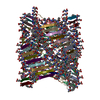
 Sample preparation
 Electron microscopy imaging
 FIELD EMISSION GUN / Accelerating voltage: 300 kV / Illumination mode: FLOOD BEAM
 Processing