Protocol: SINGLE WAVELENGTH / Monochromatic (M) / Laue (L): M / Scattering type: x-ray
Radiation wavelength
Wavelength: 0.9322 Å / Relative weight: 1
Reflection
Resolution: 2.6→99.99 Å
Reflection shell
Resolution: 2.6→2.67 Å / % possible all: 100
-
Processing
Software
Name
Version
Classification
REFMAC
5.5.0109
refinement
SCALA
datascaling
Refinement
Resolution: 2.6→99.99 Å / Cor.coef. Fo:Fc: 0.937 / Cor.coef. Fo:Fc free: 0.911 / SU B: 0.002 / SU ML: 0 / Cross valid method: THROUGHOUT / ESU R: 0.297 / ESU R Free: 0.344 / Stereochemistry target values: MAXIMUM LIKELIHOOD / Details: HYDROGENS HAVE BEEN ADDED IN THE RIDING POSITIONS
Rfactor
Num. reflection
% reflection
Selection details
Rfree
0.26783
3031
5.1 %
RANDOM
Rwork
0.23068
-
-
-
obs
0.23257
56969
99.93 %
-
Solvent computation
Ion probe radii: 0.8 Å / Shrinkage radii: 0.8 Å / VDW probe radii: 1.4 Å / Solvent model: MASK
Displacement parameters
Biso mean: 36.493 Å2
Baniso -1
Baniso -2
Baniso -3
1-
1.19 Å2
0 Å2
0.72 Å2
2-
-
-1.2 Å2
0 Å2
3-
-
-
0.15 Å2
Refinement step
Cycle: 1 / Resolution: 2.6→99.99 Å
Protein
Nucleic acid
Ligand
Solvent
Total
Num. atoms
12534
0
492
326
13352
LS refinement shell
Resolution: 2.6→2.667 Å / Total num. of bins used: 20
Rfactor
Num. reflection
% reflection
Rfree
0.377
239
-
Rwork
0.313
4174
-
obs
-
-
99.98 %
+
About Yorodumi
-
News
-
Feb 9, 2022. New format data for meta-information of EMDB entries
New format data for meta-information of EMDB entries
Version 3 of the EMDB header file is now the official format.
The previous official version 1.9 will be removed from the archive.
In the structure databanks used in Yorodumi, some data are registered as the other names, "COVID-19 virus" and "2019-nCoV". Here are the details of the virus and the list of structure data.
Jan 31, 2019. EMDB accession codes are about to change! (news from PDBe EMDB page)
EMDB accession codes are about to change! (news from PDBe EMDB page)
The allocation of 4 digits for EMDB accession codes will soon come to an end. Whilst these codes will remain in use, new EMDB accession codes will include an additional digit and will expand incrementally as the available range of codes is exhausted. The current 4-digit format prefixed with “EMD-” (i.e. EMD-XXXX) will advance to a 5-digit format (i.e. EMD-XXXXX), and so on. It is currently estimated that the 4-digit codes will be depleted around Spring 2019, at which point the 5-digit format will come into force.
The EM Navigator/Yorodumi systems omit the EMD- prefix.
Related info.:Q: What is EMD? / ID/Accession-code notation in Yorodumi/EM Navigator
Yorodumi is a browser for structure data from EMDB, PDB, SASBDB, etc.
This page is also the successor to EM Navigator detail page, and also detail information page/front-end page for Omokage search.
The word "yorodu" (or yorozu) is an old Japanese word meaning "ten thousand". "mi" (miru) is to see.
Related info.:EMDB / PDB / SASBDB / Comparison of 3 databanks / Yorodumi Search / Aug 31, 2016. New EM Navigator & Yorodumi / Yorodumi Papers / Jmol/JSmol / Function and homology information / Changes in new EM Navigator and Yorodumi
 Movie
Movie Controller

 Yorodumi
 Open data
 Basic information
 Components
 Keywords
 Function and homology information
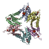
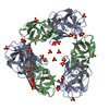
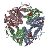
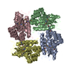
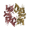
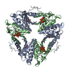
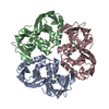
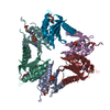
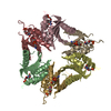
 Cyanophage S-TIM5 (virus)
 X-RAY DIFFRACTION /
  Authors
 Citation
 Structure visualization
 Downloads & links
 Other downloads


 PDBj
PDBj Assembly
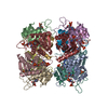


 Mass: 65.409 Da / Num. of mol.: 12 / Source method: obtained synthetically / Formula: Zn
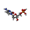
 Mass: 307.197 Da / Num. of mol.: 12 / Source method: obtained synthetically / Formula: C9H14N3O7P
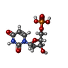
 Type: DNA linking / Mass: 308.182 Da / Num. of mol.: 12 / Source method: obtained synthetically / Formula: C9H13N2O8P
 Mass: 18.015 Da / Num. of mol.: 326 / Source method: isolated from a natural source / Formula: H2O
 Sample preparation
 / Beamline: ID14-4 / Wavelength: 0.9322 Å
 Processing