 Movie
Movie Controller


+ Open data

- Basic information
| Entry | Database: PDB / ID: 7lq6 | ||||||
|---|---|---|---|---|---|---|---|
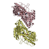
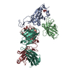
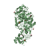
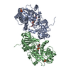
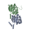
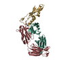
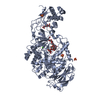
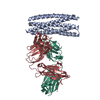
| Title | CryoEM structure of Escherichia coli PBP1b | ||||||
 Components Components | Penicillin-binding protein 1B | ||||||
 Keywords Keywords | TRANSFERASE / HYDROLASE / Penicillin binding protein / glycosyltransferase / transpeptidase | ||||||
| Function / homology |  Function and homology information Function and homology informationpositive regulation of bipolar cell growth / cell wall repair / peptidoglycan glycosyltransferase / Action of antimicrobials / peptidoglycan glycosyltransferase activity / serine-type D-Ala-D-Ala carboxypeptidase / serine-type D-Ala-D-Ala carboxypeptidase activity / penicillin binding / peptidoglycan biosynthetic process / peptidoglycan-based cell wall ...positive regulation of bipolar cell growth / cell wall repair / peptidoglycan glycosyltransferase / Action of antimicrobials / peptidoglycan glycosyltransferase activity / serine-type D-Ala-D-Ala carboxypeptidase / serine-type D-Ala-D-Ala carboxypeptidase activity / penicillin binding / peptidoglycan biosynthetic process / peptidoglycan-based cell wall / regulation of cell shape / outer membrane-bounded periplasmic space / response to antibiotic / proteolysis / membrane / plasma membrane Similarity search - Function | ||||||
| Biological species |  | ||||||
| Method | ELECTRON MICROSCOPY / single particle reconstruction / cryo EM / Resolution: 3.28 Å | ||||||
 Authors Authors | Caveney, N.A. / Workman, S.D. / Yan, R. / Atkinson, C.E. / Yu, Z. / Strynadka, N.C.J. | ||||||
| Funding support |  Canada, 1items Canada, 1items
| ||||||
 Citation Citation | Journal: Nat Commun / Year: 2021 Title: CryoEM structure of the antibacterial target PBP1b at 3.3 Å resolution. Authors: Nathanael A Caveney / Sean D Workman / Rui Yan / Claire E Atkinson / Zhiheng Yu / Natalie C J Strynadka /  Abstract: The pathway for the biosynthesis of the bacterial cell wall is one of the most prolific antibiotic targets, exemplified by the widespread use of β-lactam antibiotics. Despite this, our structural ...The pathway for the biosynthesis of the bacterial cell wall is one of the most prolific antibiotic targets, exemplified by the widespread use of β-lactam antibiotics. Despite this, our structural understanding of class A penicillin binding proteins, which perform the last two steps in this pathway, is incomplete due to the inherent difficulty in their crystallization and the complexity of their substrates. Here, we determine the near atomic resolution structure of the 83 kDa class A PBP from Escherichia coli, PBP1b, using cryogenic electron microscopy and a styrene maleic acid anhydride membrane mimetic. PBP1b, in its apo form, is seen to exhibit a distinct conformation in comparison to Moenomycin-bound crystal structures. The work herein paves the way for the use of cryoEM in structure-guided antibiotic development for this notoriously difficult to crystalize class of proteins and their complex substrates. | ||||||
| History |
|
- Structure visualization
| Movie |
Movie viewer |
|---|---|
| Structure viewer | Molecule: MolmilJmol/JSmol |
- Downloads & links
-Download
| PDBx/mmCIF format | 7lq6.cif.gz | 128.7 KB | Display | PDBx/mmCIF format |
|---|---|---|---|---|
| PDB format | pdb7lq6.ent.gz | 98.2 KB | Display | PDB format |
| PDBx/mmJSON format | 7lq6.json.gz | Tree view | PDBx/mmJSON format | |
| Others |  Other downloads Other downloads |
-Validation report
| Arichive directory | https://data.pdbj.org/pub/pdb/validation_reports/lq/7lq6ftp://data.pdbj.org/pub/pdb/validation_reports/lq/7lq6 | HTTPS FTP |
|---|
-Related structure data
| Related structure data |  23482MC M: map data used to model this data C: citing same article ( |
|---|---|
| Similar structure data |


-Links
 PDBj
PDBj
- Assembly
| Deposited unit | 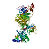
|
|---|---|
| 1 |
|
-Components
| #1: Protein | Mass: 83280.352 Da / Num. of mol.: 1 / Fragment: UNP residues 58-804 Source method: isolated from a genetically manipulated source Source: (gene. exp.) Strain: K12 / Gene: mrcB, pbpF, ponB, b0149, JW0145 / Production host: References: UniProt: P02919, peptidoglycan glycosyltransferase, serine-type D-Ala-D-Ala carboxypeptidase |
|---|
-Experimental details
-Experiment
| Experiment | Method: ELECTRON MICROSCOPY |
|---|---|
| EM experiment | Aggregation state: PARTICLE / 3D reconstruction method: single particle reconstruction |
- Sample preparation
| Component | Name: Escherichia coli PBP1b in SMA / Type: COMPLEX / Entity ID: all / Source: RECOMBINANT |
|---|---|
| Molecular weight | Value: 0.083 MDa / Experimental value: NO |
| Source (natural) | Organism: |
| Source (recombinant) | Organism: |
| Buffer solution | pH: 8 |
| Specimen | Conc.: 0.125 mg/ml / Embedding applied: NO / Shadowing applied: NO / Staining applied: NO / Vitrification applied: YES |
| Specimen support | Grid material: COPPER / Grid mesh size: 300 divisions/in. / Grid type: Quantifoil R1.2/1.3 |
| Vitrification | Instrument: FEI VITROBOT MARK IV / Cryogen name: ETHANE / Humidity: 100 % / Chamber temperature: 277.15 K / Details: 3 blot force, 3 second blot |
- Electron microscopy imaging
| Experimental equipment |  Model: Titan Krios / Image courtesy: FEI Company |
|---|---|
| Microscopy | Model: TFS KRIOS |
| Electron gun | Electron source:  FIELD EMISSION GUN / Accelerating voltage: 300 kV / Illumination mode: OTHER FIELD EMISSION GUN / Accelerating voltage: 300 kV / Illumination mode: OTHER |
| Electron lens | Mode: OTHER / Calibrated magnification: 105000 X / Cs: 2.7 mm |
| Specimen holder | Cryogen: NITROGEN / Specimen holder model: FEI TITAN KRIOS AUTOGRID HOLDER |
| Image recording | Electron dose: 60 e/Å2 / Film or detector model: GATAN K3 (6k x 4k) |
- Processing
| EM software |
| |||||||||
|---|---|---|---|---|---|---|---|---|---|---|
| CTF correction | Type: PHASE FLIPPING AND AMPLITUDE CORRECTION | |||||||||
| 3D reconstruction | Resolution: 3.28 Å / Resolution method: FSC 0.143 CUT-OFF / Num. of particles: 462997 / Symmetry type: POINT |
