 Movie
Movie Controller


+ Open data

- Basic information
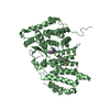
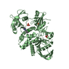
| Entry | Database: PDB / ID: 7lq4 | ||||||
|---|---|---|---|---|---|---|---|
| Title | Rr (RsiG)2-(c-di-GMP)2-WhiG complex | ||||||
 Components Components |
| ||||||
 Keywords Keywords | TRANSCRIPTION / RsiG / WhiG / Rubrobacter radiotolerans / c-di-GMP / sigma / anti-sigma / evolution / coiled coil | ||||||
| Function / homology | : / RsiG-like / nucleotide binding / metal ion binding / Chem-C2E / RsiG-like domain-containing protein Function and homology information Function and homology information | ||||||
| Biological species |  Rubrobacter radiotolerans (bacteria) Rubrobacter radiotolerans (bacteria) | ||||||
| Method |  X-RAY DIFFRACTION / SYNCHROTRON / MOLECULAR REPLACEMENT / molecular replacement / Resolution: 2.9 Å X-RAY DIFFRACTION / SYNCHROTRON / MOLECULAR REPLACEMENT / molecular replacement / Resolution: 2.9 Å | ||||||
 Authors Authors | Schumacher, M.A. / Brennan, R.G. | ||||||
| Funding support |  United States, 1items United States, 1items
| ||||||
 Citation Citation | Journal: Proc.Natl.Acad.Sci.USA / Year: 2021 Title: Evolution of a sigma-(c-di-GMP)-anti-sigma switch. Authors: Schumacher, M.A. / Gallagher, K.A. / Holmes, N.A. / Chandra, G. / Henderson, M. / Kysela, D.T. / Brennan, R.G. / Buttner, M.J. | ||||||
| History |
|
- Structure visualization
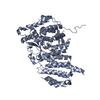
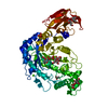
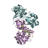
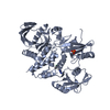
| Structure viewer | Molecule: MolmilJmol/JSmol |
|---|
- Downloads & links
-Download
| PDBx/mmCIF format | 7lq4.cif.gz | 156.7 KB | Display | PDBx/mmCIF format |
|---|---|---|---|---|
| PDB format | pdb7lq4.ent.gz | 121.5 KB | Display | PDB format |
| PDBx/mmJSON format | 7lq4.json.gz | Tree view | PDBx/mmJSON format | |
| Others |  Other downloads Other downloads |
-Validation report
| Arichive directory | https://data.pdbj.org/pub/pdb/validation_reports/lq/7lq4ftp://data.pdbj.org/pub/pdb/validation_reports/lq/7lq4 | HTTPS FTP |
|---|
-Related structure data
| Related structure data | 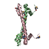 7lq2C 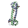 7lq3C  6pfjS C: citing same article ( S: Starting model for refinement |
|---|---|
| Similar structure data |


-Links
 PDBj
PDBj
- Assembly
| Deposited unit | 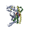
| ||||||||
|---|---|---|---|---|---|---|---|---|---|
| 1 |
| ||||||||
| Unit cell |
|
-Components
| #1: Protein | Mass: 12955.107 Da / Num. of mol.: 2 Source method: isolated from a genetically manipulated source Source: (gene. exp.) Rubrobacter radiotolerans (bacteria) / Gene: RradSPS_1442 / Production host: #2: Protein | | Mass: 22183.393 Da / Num. of mol.: 1 Source method: isolated from a genetically manipulated source Source: (gene. exp.) Rubrobacter radiotolerans (bacteria) / Production host: #3: Chemical | 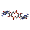  Mass: 690.411 Da / Num. of mol.: 2 / Source method: obtained synthetically / Formula: C20H24N10O14P2 Mass: 690.411 Da / Num. of mol.: 2 / Source method: obtained synthetically / Formula: C20H24N10O14P2#4: Water | ChemComp-HOH / |  Mass: 18.015 Da / Num. of mol.: 11 / Source method: isolated from a natural source / Formula: H2O Mass: 18.015 Da / Num. of mol.: 11 / Source method: isolated from a natural source / Formula: H2OHas ligand of interest | N | Sequence details | The complete sequence of WhiG is VRVSIERLWSQYFEARAKLGSLEPDEREAAETLEKRVRGLKDRLVVNYSPLVKYAAGRVT ...The complete sequence of WhiG is VRVSIERLWS | |
|---|
-Experimental details
-Experiment
| Experiment | Method: X-RAY DIFFRACTION / Number of used crystals: 1 |
|---|
- Sample preparation
| Crystal | Density Matthews: 2.26 Å3/Da / Density % sol: 45.55 % |
|---|---|
| Crystal grow | Temperature: 298 K / Method: vapor diffusion, hanging drop / Details: 0.1 M Tris pH 8.0, 0.2 M MgCl2, 23% PEG 3350 |
-Data collection
| Diffraction | Mean temperature: 100 K / Serial crystal experiment: N |
|---|---|
| Diffraction source | Source: SYNCHROTRON / Site: ALS / Beamline: 8.3.1 / Wavelength: 1.01 Å |
| Detector | Type: DECTRIS PILATUS 6M / Detector: PIXEL / Date: Jan 23, 2020 |
| Radiation | Protocol: SINGLE WAVELENGTH / Monochromatic (M) / Laue (L): M / Scattering type: x-ray |
| Radiation wavelength | Wavelength: 1.01 Å / Relative weight: 1 |
| Reflection | Resolution: 2.9→66.48 Å / Num. obs: 9237 / % possible obs: 91.9 % / Redundancy: 2.9 % / Biso Wilson estimate: 81.78 Å2 / CC1/2: 0.999 / Rpim(I) all: 0.035 / Rsym value: 0.043 / Net I/σ(I): 17.1 |
| Reflection shell | Resolution: 2.93→3.04 Å / Num. unique obs: 601 / CC1/2: 0.804 / Rpim(I) all: 0.23 / Rsym value: 0.24 |
-Phasing
| Phasing | Method: molecular replacement |
|---|
- Processing
| Software |
| ||||||||||||||||||||||||||||||||||||||||
|---|---|---|---|---|---|---|---|---|---|---|---|---|---|---|---|---|---|---|---|---|---|---|---|---|---|---|---|---|---|---|---|---|---|---|---|---|---|---|---|---|---|
| Refinement | Method to determine structure: MOLECULAR REPLACEMENT Starting model: 6PFJ Resolution: 2.9→66.48 Å / SU ML: 0.32 / Cross valid method: THROUGHOUT / σ(F): 1.36 / Phase error: 32.63 / Stereochemistry target values: ML
| ||||||||||||||||||||||||||||||||||||||||
| Solvent computation | Shrinkage radii: 0.72 Å / VDW probe radii: 1 Å / Solvent model: FLAT BULK SOLVENT MODEL / Bsol: 80.018 Å2 / ksol: 0.346 e/Å3 | ||||||||||||||||||||||||||||||||||||||||
| Displacement parameters | Biso max: 223.53 Å2 / Biso mean: 106.07 Å2 / Biso min: 38.43 Å2
| ||||||||||||||||||||||||||||||||||||||||
| Refinement step | Cycle: final / Resolution: 2.9→66.48 Å
| ||||||||||||||||||||||||||||||||||||||||
| Refine LS restraints |
| ||||||||||||||||||||||||||||||||||||||||
| LS refinement shell | Refine-ID: X-RAY DIFFRACTION / Rfactor Rfree error: 0
| ||||||||||||||||||||||||||||||||||||||||
| Refinement TLS params. | Method: refined / Origin x: -17.0899 Å / Origin y: -4.8801 Å / Origin z: -24.5724 Å
| ||||||||||||||||||||||||||||||||||||||||
| Refinement TLS group |
|
