 Movie
Movie Controller


+ Open data

- Basic information
| Entry | Database: PDB / ID: 7lq2 | ||||||
|---|---|---|---|---|---|---|---|
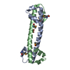
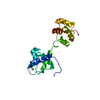
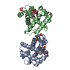
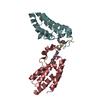
| Title | Apo Rr RsiG- crystal form 1 | ||||||
 Components Components | RR RsiG | ||||||
 Keywords Keywords | TRANSCRIPTION / RsiG / single-motif / WhiG / evolution / homodimer / Rubrobacter radiotolerans | ||||||
| Function / homology | : / RsiG-like / nucleotide binding / metal ion binding / ISOPROPYL ALCOHOL / RsiG-like domain-containing protein Function and homology information Function and homology information | ||||||
| Biological species |  Rubrobacter radiotolerans (bacteria) Rubrobacter radiotolerans (bacteria) | ||||||
| Method |  X-RAY DIFFRACTION / SYNCHROTRON / SAD / Resolution: 1.85 Å X-RAY DIFFRACTION / SYNCHROTRON / SAD / Resolution: 1.85 Å | ||||||
 Authors Authors | Schumacher, M.A. / Brennan, R.G. | ||||||
| Funding support |  United States, 1items United States, 1items
| ||||||
 Citation Citation | Journal: Proc.Natl.Acad.Sci.USA / Year: 2021 Title: Evolution of a sigma-(c-di-GMP)-anti-sigma switch. Authors: Schumacher, M.A. / Gallagher, K.A. / Holmes, N.A. / Chandra, G. / Henderson, M. / Kysela, D.T. / Brennan, R.G. / Buttner, M.J. | ||||||
| History |
|
- Structure visualization
| Structure viewer | Molecule: MolmilJmol/JSmol |
|---|
- Downloads & links
-Download
| PDBx/mmCIF format | 7lq2.cif.gz | 198.8 KB | Display | PDBx/mmCIF format |
|---|---|---|---|---|
| PDB format | pdb7lq2.ent.gz | 161.4 KB | Display | PDB format |
| PDBx/mmJSON format | 7lq2.json.gz | Tree view | PDBx/mmJSON format | |
| Others |  Other downloads Other downloads |
-Validation report
| Arichive directory | https://data.pdbj.org/pub/pdb/validation_reports/lq/7lq2ftp://data.pdbj.org/pub/pdb/validation_reports/lq/7lq2 | HTTPS FTP |
|---|
-Related structure data


-Links
 PDBj
PDBj
- Assembly
| Deposited unit | 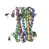
| ||||||||
|---|---|---|---|---|---|---|---|---|---|
| 1 | 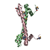
| ||||||||
| 2 | 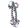
| ||||||||
| 3 | 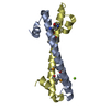
| ||||||||
| Unit cell |
|
-Components
| #1: Protein | Mass: 9679.238 Da / Num. of mol.: 6 Source method: isolated from a genetically manipulated source Source: (gene. exp.) Rubrobacter radiotolerans (bacteria) / Gene: RradSPS_1442 / Production host: #2: Chemical |   Mass: 24.305 Da / Num. of mol.: 2 / Source method: obtained synthetically / Formula: Mg / Feature type: SUBJECT OF INVESTIGATION Mass: 24.305 Da / Num. of mol.: 2 / Source method: obtained synthetically / Formula: Mg / Feature type: SUBJECT OF INVESTIGATION#3: Chemical |   Mass: 60.095 Da / Num. of mol.: 2 / Source method: obtained synthetically / Formula: C3H8O / Feature type: SUBJECT OF INVESTIGATION / Comment: alkaloid*YM Mass: 60.095 Da / Num. of mol.: 2 / Source method: obtained synthetically / Formula: C3H8O / Feature type: SUBJECT OF INVESTIGATION / Comment: alkaloid*YM#4: Water | ChemComp-HOH / |  Mass: 18.015 Da / Num. of mol.: 443 / Source method: isolated from a natural source / Formula: H2O Mass: 18.015 Da / Num. of mol.: 443 / Source method: isolated from a natural source / Formula: H2OHas ligand of interest | Y | Has protein modification | Y | |
|---|
-Experimental details
-Experiment
| Experiment | Method: X-RAY DIFFRACTION / Number of used crystals: 1 |
|---|
- Sample preparation
| Crystal | Density Matthews: 2.89 Å3/Da / Density % sol: 57.38 % |
|---|---|
| Crystal grow | Temperature: 298 K / Method: vapor diffusion, hanging drop / Details: 12% isopropanol, 0.1 M HEPES pH 7.5, 0.1 M MgCl2 |
-Data collection
| Diffraction | Mean temperature: 100 K / Serial crystal experiment: N |
|---|---|
| Diffraction source | Source: SYNCHROTRON / Site: ALS / Beamline: 5.0.2 / Wavelength: 0.979 Å |
| Detector | Type: DECTRIS PILATUS 6M / Detector: PIXEL / Date: Jun 27, 2020 |
| Radiation | Protocol: SINGLE WAVELENGTH / Monochromatic (M) / Laue (L): M / Scattering type: x-ray |
| Radiation wavelength | Wavelength: 0.979 Å / Relative weight: 1 |
| Reflection | Resolution: 1.85→50.46 Å / Num. obs: 57371 / % possible obs: 99.6 % / Redundancy: 6.6 % / CC1/2: 0.998 / Rpim(I) all: 0.032 / Rsym value: 0.053 / Net I/σ(I): 17.8 |
| Reflection shell | Resolution: 1.85→1.92 Å / Mean I/σ(I) obs: 2.4 / Num. unique obs: 5313 / CC1/2: 0.898 / Rpim(I) all: 0.374 / Rsym value: 0.464 |
-Phasing
| Phasing | Method: SAD |
|---|
- Processing
| Software |
| ||||||||||||||||||||||||||||||||||||||||||||||||||||||||||||||||||||||||||||||
|---|---|---|---|---|---|---|---|---|---|---|---|---|---|---|---|---|---|---|---|---|---|---|---|---|---|---|---|---|---|---|---|---|---|---|---|---|---|---|---|---|---|---|---|---|---|---|---|---|---|---|---|---|---|---|---|---|---|---|---|---|---|---|---|---|---|---|---|---|---|---|---|---|---|---|---|---|---|---|---|
| Refinement | Method to determine structure: SAD / Resolution: 1.85→50.457 Å / SU ML: 0.16 / Cross valid method: THROUGHOUT / σ(F): 0 / Phase error: 20.98 / Stereochemistry target values: ML
| ||||||||||||||||||||||||||||||||||||||||||||||||||||||||||||||||||||||||||||||
| Solvent computation | Shrinkage radii: 0.9 Å / VDW probe radii: 1.11 Å / Solvent model: FLAT BULK SOLVENT MODEL | ||||||||||||||||||||||||||||||||||||||||||||||||||||||||||||||||||||||||||||||
| Displacement parameters | Biso max: 123.01 Å2 / Biso mean: 28.1805 Å2 / Biso min: 10.33 Å2 | ||||||||||||||||||||||||||||||||||||||||||||||||||||||||||||||||||||||||||||||
| Refinement step | Cycle: final / Resolution: 1.85→50.457 Å
| ||||||||||||||||||||||||||||||||||||||||||||||||||||||||||||||||||||||||||||||
| LS refinement shell | Refine-ID: X-RAY DIFFRACTION / Rfactor Rfree error: 0
| ||||||||||||||||||||||||||||||||||||||||||||||||||||||||||||||||||||||||||||||
| Refinement TLS params. | Method: refined / Origin x: 44.5479 Å / Origin y: 22.0469 Å / Origin z: 66.4727 Å
| ||||||||||||||||||||||||||||||||||||||||||||||||||||||||||||||||||||||||||||||
| Refinement TLS group |
|
