 Movie
Movie Controller

[English] 日本語
 Yorodumi
- PDB-7lpz: Crystal structures and ribonuclease activity of the Flavivirus ho... -
+ Open data

- Basic information
| Entry | Database: PDB / ID: 7lpz | ||||||||||||
|---|---|---|---|---|---|---|---|---|---|---|---|---|---|
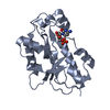
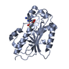
| Title | Crystal structures and ribonuclease activity of the Flavivirus host factor ERI3 that is involved in viral RNA synthesis define the ERI subfamily of structure-specific 3-prime - 5-prime exoribonucleases | ||||||||||||
 Components Components | ERI1 exoribonuclease 3 | ||||||||||||
 Keywords Keywords | HYDROLASE / HYDROLASE METAL BINDING | ||||||||||||
| Function / homology |  Function and homology information Function and homology information3'-5'-RNA exonuclease activity / Hydrolases; Acting on ester bonds / RNA binding / metal ion binding Similarity search - Function | ||||||||||||
| Biological species |  Homo sapiens (human) Homo sapiens (human) | ||||||||||||
| Method |  X-RAY DIFFRACTION / SYNCHROTRON / MOLECULAR REPLACEMENT / Resolution: 1.55 Å X-RAY DIFFRACTION / SYNCHROTRON / MOLECULAR REPLACEMENT / Resolution: 1.55 Å | ||||||||||||
 Authors Authors | Thapar, R. / Andrew, A.S. / Tainer, J.A. | ||||||||||||
| Funding support |  United States, 3items United States, 3items
| ||||||||||||
 Citation Citation | Journal: To Be Published Title: Crystal structures and ribonuclease activity of the Flavivirus host factor ERI3 that is involved in viral RNA synthesis define the ERI subfamily of structure-specific 3' - 5' exoribonucleases Authors: Thapar, R. / Arvai, A.S. / Tainer, J.A. | ||||||||||||
| History |
|
- Structure visualization
| Structure viewer | Molecule: MolmilJmol/JSmol |
|---|
- Downloads & links
-Download
| PDBx/mmCIF format | 7lpz.cif.gz | 233.2 KB | Display | PDBx/mmCIF format |
|---|---|---|---|---|
| PDB format | pdb7lpz.ent.gz | 155.6 KB | Display | PDB format |
| PDBx/mmJSON format | 7lpz.json.gz | Tree view | PDBx/mmJSON format | |
| Others |  Other downloads Other downloads |
-Validation report
| Arichive directory | https://data.pdbj.org/pub/pdb/validation_reports/lp/7lpzftp://data.pdbj.org/pub/pdb/validation_reports/lp/7lpz | HTTPS FTP |
|---|
-Related structure data
| Related structure data |  7k05C 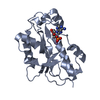 7lpyC 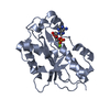 7lq0C 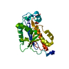 2xriS C: citing same article ( S: Starting model for refinement |
|---|---|
| Similar structure data |


-Links
 PDBj
PDBj


- Assembly
| Deposited unit | 
| ||||||||||||||||||||||||||||||||||||||||||||||||||||||||||||||||||||||||||||||||||||||
|---|---|---|---|---|---|---|---|---|---|---|---|---|---|---|---|---|---|---|---|---|---|---|---|---|---|---|---|---|---|---|---|---|---|---|---|---|---|---|---|---|---|---|---|---|---|---|---|---|---|---|---|---|---|---|---|---|---|---|---|---|---|---|---|---|---|---|---|---|---|---|---|---|---|---|---|---|---|---|---|---|---|---|---|---|---|---|---|
| 1 | 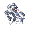
| ||||||||||||||||||||||||||||||||||||||||||||||||||||||||||||||||||||||||||||||||||||||
| 2 | 
| ||||||||||||||||||||||||||||||||||||||||||||||||||||||||||||||||||||||||||||||||||||||
| Unit cell |
| ||||||||||||||||||||||||||||||||||||||||||||||||||||||||||||||||||||||||||||||||||||||
| Noncrystallographic symmetry (NCS) | NCS domain:
NCS domain segments:
NCS oper: (Code: givenMatrix: (0.0120605054072, 0.115438158984, 0.993241448823), (0.205984769455, 0.971722321892, -0.115438312049), (-0.978480873069, 0.205984855235, -0.0120590402472)Vector: 47. ...NCS oper: (Code: given Matrix: (0.0120605054072, 0.115438158984, 0.993241448823), Vector: |
-Components
| #1: Protein | Mass: 25068.066 Da / Num. of mol.: 2 Source method: isolated from a genetically manipulated source Source: (gene. exp.) Homo sapiens (human) / Gene: ERI3, PINT1, PRNPIP, PRNPIP1 / Production host:  References: UniProt: O43414, Hydrolases; Acting on ester bonds #2: Chemical |   Mass: 347.221 Da / Num. of mol.: 2 / Source method: obtained synthetically / Formula: C10H14N5O7P / Feature type: SUBJECT OF INVESTIGATION / Comment: AMP*YM Mass: 347.221 Da / Num. of mol.: 2 / Source method: obtained synthetically / Formula: C10H14N5O7P / Feature type: SUBJECT OF INVESTIGATION / Comment: AMP*YM#3: Chemical | ChemComp-SM /   Mass: 150.360 Da / Num. of mol.: 4 / Source method: obtained synthetically / Formula: Sm / Feature type: SUBJECT OF INVESTIGATION Mass: 150.360 Da / Num. of mol.: 4 / Source method: obtained synthetically / Formula: Sm / Feature type: SUBJECT OF INVESTIGATION#4: Chemical | ChemComp-GOL / |   Mass: 92.094 Da / Num. of mol.: 1 / Source method: obtained synthetically / Formula: C3H8O3 Mass: 92.094 Da / Num. of mol.: 1 / Source method: obtained synthetically / Formula: C3H8O3#5: Water | ChemComp-HOH / |  Mass: 18.015 Da / Num. of mol.: 329 / Source method: isolated from a natural source / Formula: H2O Mass: 18.015 Da / Num. of mol.: 329 / Source method: isolated from a natural source / Formula: H2OHas ligand of interest | Y | Has protein modification | Y | |
|---|
-Experimental details
-Experiment
| Experiment | Method: X-RAY DIFFRACTION / Number of used crystals: 1 |
|---|
- Sample preparation
| Crystal | Density Matthews: 2.12 Å3/Da / Density % sol: 42.03 % |
|---|---|
| Crystal grow | Temperature: 290 K / Method: vapor diffusion, hanging drop Details: 30% PEG 3350, 25% saturated KCl, 200 mM Imidazole / Malate buffer 8.0, 2.5 mM samarium (III) acetate, 18mM AMP, 3.3 mM N-{2-[4-(thiophen-2-yl)-1H-imidazol-2-yl]ethyl}-2,3-dihydro-1,4- ...Details: 30% PEG 3350, 25% saturated KCl, 200 mM Imidazole / Malate buffer 8.0, 2.5 mM samarium (III) acetate, 18mM AMP, 3.3 mM N-{2-[4-(thiophen-2-yl)-1H-imidazol-2-yl]ethyl}-2,3-dihydro-1,4-benzodioxine-2-carboxamide (from Life Chemicals) |
-Data collection
| Diffraction | Mean temperature: 100 K / Serial crystal experiment: N |
|---|---|
| Diffraction source | Source: SYNCHROTRON / Site: SSRL / Beamline: BL9-2 / Wavelength: 0.97946 Å |
| Detector | Type: DECTRIS PILATUS 6M / Detector: PIXEL / Date: Jan 11, 2021 |
| Radiation | Protocol: SINGLE WAVELENGTH / Monochromatic (M) / Laue (L): M / Scattering type: x-ray |
| Radiation wavelength | Wavelength: 0.97946 Å / Relative weight: 1 |
| Reflection | Resolution: 1.55→37.39 Å / Num. obs: 407820 / % possible obs: 99.4 % / Redundancy: 6.56 % / Biso Wilson estimate: 28.81 Å2 / CC1/2: 1 / Net I/σ(I): 22.42 |
| Reflection shell | Resolution: 1.55→1.64 Å / Num. unique obs: 63859 / CC1/2: 0.631 |
- Processing
| Software |
| |||||||||||||||||||||||||||||||||||||||||||||||||||||||||||||||||||||||||||||||||||||||||||||||||||||||||||||||||||||||||||||||||||||||||||||||||||||||||||||||||||||||||||||||
|---|---|---|---|---|---|---|---|---|---|---|---|---|---|---|---|---|---|---|---|---|---|---|---|---|---|---|---|---|---|---|---|---|---|---|---|---|---|---|---|---|---|---|---|---|---|---|---|---|---|---|---|---|---|---|---|---|---|---|---|---|---|---|---|---|---|---|---|---|---|---|---|---|---|---|---|---|---|---|---|---|---|---|---|---|---|---|---|---|---|---|---|---|---|---|---|---|---|---|---|---|---|---|---|---|---|---|---|---|---|---|---|---|---|---|---|---|---|---|---|---|---|---|---|---|---|---|---|---|---|---|---|---|---|---|---|---|---|---|---|---|---|---|---|---|---|---|---|---|---|---|---|---|---|---|---|---|---|---|---|---|---|---|---|---|---|---|---|---|---|---|---|---|---|---|---|---|
| Refinement | Method to determine structure: MOLECULAR REPLACEMENT Starting model: 2XRI Resolution: 1.55→37.39 Å / SU ML: 0.199 / Cross valid method: FREE R-VALUE / σ(F): 0 / Phase error: 25.8326 Stereochemistry target values: GeoStd + Monomer Library + CDL v1.2
| |||||||||||||||||||||||||||||||||||||||||||||||||||||||||||||||||||||||||||||||||||||||||||||||||||||||||||||||||||||||||||||||||||||||||||||||||||||||||||||||||||||||||||||||
| Solvent computation | Shrinkage radii: 0.8 Å / VDW probe radii: 1.1 Å / Solvent model: FLAT BULK SOLVENT MODEL | |||||||||||||||||||||||||||||||||||||||||||||||||||||||||||||||||||||||||||||||||||||||||||||||||||||||||||||||||||||||||||||||||||||||||||||||||||||||||||||||||||||||||||||||
| Displacement parameters | Biso mean: 44.83 Å2 | |||||||||||||||||||||||||||||||||||||||||||||||||||||||||||||||||||||||||||||||||||||||||||||||||||||||||||||||||||||||||||||||||||||||||||||||||||||||||||||||||||||||||||||||
| Refinement step | Cycle: LAST / Resolution: 1.55→37.39 Å
| |||||||||||||||||||||||||||||||||||||||||||||||||||||||||||||||||||||||||||||||||||||||||||||||||||||||||||||||||||||||||||||||||||||||||||||||||||||||||||||||||||||||||||||||
| Refine LS restraints |
| |||||||||||||||||||||||||||||||||||||||||||||||||||||||||||||||||||||||||||||||||||||||||||||||||||||||||||||||||||||||||||||||||||||||||||||||||||||||||||||||||||||||||||||||
| Refine LS restraints NCS | Type: Torsion NCS / Rms dev position: 0.533027182314 Å | |||||||||||||||||||||||||||||||||||||||||||||||||||||||||||||||||||||||||||||||||||||||||||||||||||||||||||||||||||||||||||||||||||||||||||||||||||||||||||||||||||||||||||||||
| LS refinement shell |
|
