 Movie
Movie Controller

[English] 日本語
 Yorodumi
- PDB-7lc9: Cryo-EM structure of the N-terminal alpha-synuclein truncation 41-140 -
+ Open data

- Basic information
| Entry | Database: PDB / ID: 7lc9 | |||||||||||||||||||||
|---|---|---|---|---|---|---|---|---|---|---|---|---|---|---|---|---|---|---|---|---|---|---|
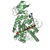
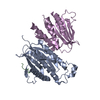
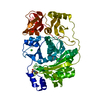
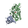
| Title | Cryo-EM structure of the N-terminal alpha-synuclein truncation 41-140 | |||||||||||||||||||||
 Components Components | Alpha-synuclein | |||||||||||||||||||||
 Keywords Keywords | PROTEIN FIBRIL / N-terminal alpha-synuclein truncation | |||||||||||||||||||||
| Function / homology |  Function and homology information Function and homology informationnegative regulation of mitochondrial electron transport, NADH to ubiquinone / : / neutral lipid metabolic process / regulation of acyl-CoA biosynthetic process / negative regulation of dopamine uptake involved in synaptic transmission / negative regulation of norepinephrine uptake / response to desipramine / positive regulation of SNARE complex assembly / positive regulation of hydrogen peroxide catabolic process / supramolecular fiber ...negative regulation of mitochondrial electron transport, NADH to ubiquinone / : / neutral lipid metabolic process / regulation of acyl-CoA biosynthetic process / negative regulation of dopamine uptake involved in synaptic transmission / negative regulation of norepinephrine uptake / response to desipramine / positive regulation of SNARE complex assembly / positive regulation of hydrogen peroxide catabolic process / supramolecular fiber / regulation of synaptic vesicle recycling / negative regulation of chaperone-mediated autophagy / mitochondrial membrane organization / regulation of reactive oxygen species biosynthetic process / positive regulation of protein localization to cell periphery / negative regulation of platelet-derived growth factor receptor signaling pathway / negative regulation of exocytosis / regulation of glutamate secretion / dopamine biosynthetic process / response to iron(II) ion / SNARE complex assembly / negative regulation of dopamine metabolic process / positive regulation of neurotransmitter secretion / regulation of macrophage activation / positive regulation of inositol phosphate biosynthetic process / regulation of norepinephrine uptake / regulation of locomotion / synaptic vesicle transport / negative regulation of microtubule polymerization / transporter regulator activity / synaptic vesicle priming / dopamine uptake involved in synaptic transmission / protein kinase inhibitor activity / regulation of dopamine secretion / negative regulation of thrombin-activated receptor signaling pathway / dynein complex binding / mitochondrial ATP synthesis coupled electron transport / positive regulation of receptor recycling / cuprous ion binding / nuclear outer membrane / response to magnesium ion / positive regulation of exocytosis / synaptic vesicle exocytosis / positive regulation of endocytosis / kinesin binding / synaptic vesicle endocytosis / enzyme inhibitor activity / cysteine-type endopeptidase inhibitor activity / response to type II interferon / negative regulation of serotonin uptake / regulation of presynapse assembly / alpha-tubulin binding / beta-tubulin binding / phospholipase binding / behavioral response to cocaine / supramolecular fiber organization / phospholipid metabolic process / cellular response to fibroblast growth factor stimulus / axon terminus / inclusion body / cellular response to epinephrine stimulus / Hsp70 protein binding / response to interleukin-1 / regulation of microtubule cytoskeleton organization / cellular response to copper ion / positive regulation of release of sequestered calcium ion into cytosol / SNARE binding / adult locomotory behavior / excitatory postsynaptic potential / protein tetramerization / phosphoprotein binding / microglial cell activation / ferrous iron binding / fatty acid metabolic process / regulation of long-term neuronal synaptic plasticity / synapse organization / PKR-mediated signaling / protein destabilization / phospholipid binding / receptor internalization / tau protein binding / long-term synaptic potentiation / terminal bouton / positive regulation of inflammatory response / synaptic vesicle membrane / actin cytoskeleton / growth cone / actin binding / cellular response to oxidative stress / neuron apoptotic process / cell cortex / histone binding / response to lipopolysaccharide / microtubule binding / chemical synaptic transmission / amyloid fibril formation / molecular adaptor activity / negative regulation of neuron apoptotic process / mitochondrial outer membrane / oxidoreductase activity Similarity search - Function | |||||||||||||||||||||
| Biological species |  Homo sapiens (human) Homo sapiens (human) | |||||||||||||||||||||
| Method | ELECTRON MICROSCOPY / helical reconstruction / cryo EM / Resolution: 3.2 Å | |||||||||||||||||||||
 Authors Authors | Xiaodan, N. / Ryan, P.M. / Jiansen, J. / Jennifer, C.L. | |||||||||||||||||||||
| Funding support |  United States, 1items United States, 1items
| |||||||||||||||||||||
 Citation Citation | Journal: Proc Natl Acad Sci U S A / Year: 2021 Title: The N terminus of α-synuclein dictates fibril formation. Authors: Ryan P McGlinchey / Xiaodan Ni / Jared A Shadish / Jiansen Jiang / Jennifer C Lee / Abstract: The generation of α-synuclein (α-syn) truncations from incomplete proteolysis plays a significant role in the pathogenesis of Parkinson's disease. It is well established that C-terminal truncations ...The generation of α-synuclein (α-syn) truncations from incomplete proteolysis plays a significant role in the pathogenesis of Parkinson's disease. It is well established that C-terminal truncations exhibit accelerated aggregation and serve as potent seeds in fibril propagation. In contrast, mechanistic understanding of N-terminal truncations remains ill defined. Previously, we found that disease-related C-terminal truncations resulted in increased fibrillar twist, accompanied by modest conformational changes in a more compact core, suggesting that the N-terminal region could be dictating fibril structure. Here, we examined three N-terminal truncations, in which deletions of 13-, 35-, and 40-residues in the N terminus modulated both aggregation kinetics and fibril morphologies. Cross-seeding experiments showed that out of the three variants, only ΔN13-α-syn (14‒140) fibrils were capable of accelerating full-length fibril formation, albeit slower than self-seeding. Interestingly, the reversed cross-seeding reactions with full-length seeds efficiently promoted all but ΔN40-α-syn (41-140). This behavior can be explained by the unique fibril structure that is adopted by 41-140 with two asymmetric protofilaments, which was determined by cryogenic electron microscopy. One protofilament resembles the previously characterized bent β-arch kernel, comprised of residues E46‒K96, whereas in the other protofilament, fewer residues (E61‒D98) are found, adopting an extended β-hairpin conformation that does not resemble other reported structures. An interfilament interface exists between residues K60‒F94 and Q62‒I88 with an intermolecular salt bridge between K80 and E83. Together, these results demonstrate a vital role for the N-terminal residues in α-syn fibril formation and structure, offering insights into the interplay of α-syn and its truncations. | |||||||||||||||||||||
| History |
|
- Structure visualization
| Movie |
Movie viewer |
|---|---|
| Structure viewer | Molecule: MolmilJmol/JSmol |
- Downloads & links
-Download
| PDBx/mmCIF format | 7lc9.cif.gz | 93.4 KB | Display | PDBx/mmCIF format |
|---|---|---|---|---|
| PDB format | pdb7lc9.ent.gz | 68.8 KB | Display | PDB format |
| PDBx/mmJSON format | 7lc9.json.gz | Tree view | PDBx/mmJSON format | |
| Others |  Other downloads Other downloads |
-Validation report
| Summary document | 7lc9_validation.pdf.gz | 712.3 KB | Display | wwPDB validaton report |
|---|---|---|---|---|
| Full document | 7lc9_full_validation.pdf.gz | 732.2 KB | Display | |
| Data in XML | 7lc9_validation.xml.gz | 21.8 KB | Display | |
| Data in CIF | 7lc9_validation.cif.gz | 32.8 KB | Display | |
| Arichive directory | https://data.pdbj.org/pub/pdb/validation_reports/lc/7lc9ftp://data.pdbj.org/pub/pdb/validation_reports/lc/7lc9 | HTTPS FTP |
-Related structure data
| Related structure data | 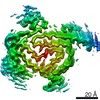 23270MC M: map data used to model this data C: citing same article ( |
|---|---|
| Similar structure data |


-Links
 PDBj
PDBj
- Assembly
| Deposited unit | 
|
|---|---|
| 1 |
|
-Components
| #1: Protein | Mass: 10346.242 Da / Num. of mol.: 12 Source method: isolated from a genetically manipulated source Source: (gene. exp.) Homo sapiens (human) / Gene: SNCA, NACP, PARK1 / Production host:  Has protein modification | N | |
|---|
-Experimental details
-Experiment
| Experiment | Method: ELECTRON MICROSCOPY |
|---|---|
| EM experiment | Aggregation state: FILAMENT / 3D reconstruction method: helical reconstruction |
- Sample preparation
| Component | Name: N-terminal alpha-synuclein truncation 41-140 / Type: COMPLEX / Entity ID: all / Source: RECOMBINANT |
|---|---|
| Source (natural) | Organism: Homo sapiens (human) |
| Source (recombinant) | Organism: |
| Buffer solution | pH: 7.4 |
| Specimen | Embedding applied: NO / Shadowing applied: NO / Staining applied: NO / Vitrification applied: YES |
| Vitrification | Cryogen name: ETHANE |
- Electron microscopy imaging
| Experimental equipment |  Model: Titan Krios / Image courtesy: FEI Company |
|---|---|
| Microscopy | Model: FEI TITAN KRIOS |
| Electron gun | Electron source:  FIELD EMISSION GUN / Accelerating voltage: 300 kV / Illumination mode: FLOOD BEAM FIELD EMISSION GUN / Accelerating voltage: 300 kV / Illumination mode: FLOOD BEAM |
| Electron lens | Mode: BRIGHT FIELD |
| Image recording | Electron dose: 55 e/Å2 / Film or detector model: GATAN K2 SUMMIT (4k x 4k) |
- Processing
| Software | Name: PHENIX / Version: 1.17.1_3660: / Classification: refinement | ||||||||||||||||||||||||
|---|---|---|---|---|---|---|---|---|---|---|---|---|---|---|---|---|---|---|---|---|---|---|---|---|---|
| EM software | Name: PHENIX / Category: model refinement | ||||||||||||||||||||||||
| CTF correction | Type: PHASE FLIPPING ONLY | ||||||||||||||||||||||||
| Helical symmerty | Angular rotation/subunit: -1.64 ° / Axial rise/subunit: 4.8 Å / Axial symmetry: C1 | ||||||||||||||||||||||||
| 3D reconstruction | Resolution: 3.2 Å / Resolution method: FSC 0.143 CUT-OFF / Num. of particles: 20867 / Symmetry type: HELICAL | ||||||||||||||||||||||||
| Refine LS restraints |
|
