 Movie
Movie Controller

[English] 日本語
 Yorodumi
- PDB-3nyb: Structure and function of the polymerase core of TRAMP, a RNA sur... -
+ Open data

- Basic information
| Entry | Database: PDB / ID: 3nyb | ||||||
|---|---|---|---|---|---|---|---|
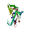
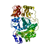
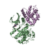
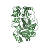
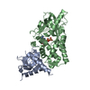
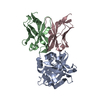
| Title | Structure and function of the polymerase core of TRAMP, a RNA surveillance complex | ||||||
 Components Components |
| ||||||
 Keywords Keywords | TRANSFERASE/RNA BINDING PROTEIN / polyA RNA polymerase / zinc knuckle protein / RNA surveillance / Mtr4p binds to Trf4p/Air2p heterodimer / TRANSFERASE-RNA BINDING PROTEIN complex | ||||||
| Function / homology |  Function and homology information Function and homology informationpolyadenylation-dependent RNA catabolic process / polyadenylation-dependent ncRNA catabolic process / polyadenylation-dependent mRNA catabolic process / TRAMP complex / nuclear mRNA surveillance of mRNA 3'-end processing / nuclear polyadenylation-dependent antisense transcript catabolic process / nuclear polyadenylation-dependent snoRNA catabolic process / nuclear polyadenylation-dependent snRNA catabolic process / meiotic DNA double-strand break formation / nuclear polyadenylation-dependent mRNA catabolic process ...polyadenylation-dependent RNA catabolic process / polyadenylation-dependent ncRNA catabolic process / polyadenylation-dependent mRNA catabolic process / TRAMP complex / nuclear mRNA surveillance of mRNA 3'-end processing / nuclear polyadenylation-dependent antisense transcript catabolic process / nuclear polyadenylation-dependent snoRNA catabolic process / nuclear polyadenylation-dependent snRNA catabolic process / meiotic DNA double-strand break formation / nuclear polyadenylation-dependent mRNA catabolic process / nuclear polyadenylation-dependent CUT catabolic process / TRAMP-dependent tRNA surveillance pathway / RNA fragment catabolic process / U4 snRNA 3'-end processing / nuclear polyadenylation-dependent rRNA catabolic process / poly(A)-dependent snoRNA 3'-end processing / RNA 3'-end processing / histone mRNA catabolic process / tRNA modification / negative regulation of DNA recombination / polynucleotide adenylyltransferase / poly(A) RNA polymerase activity / 5'-deoxyribose-5-phosphate lyase activity / base-excision repair / molecular adaptor activity / protein-macromolecule adaptor activity / cell division / mRNA binding / nucleolus / RNA binding / zinc ion binding / ATP binding / metal ion binding / nucleus / cytosol Similarity search - Function | ||||||
| Biological species |  | ||||||
| Method |  X-RAY DIFFRACTION / SYNCHROTRON / SAD / Resolution: 2.7007 Å X-RAY DIFFRACTION / SYNCHROTRON / SAD / Resolution: 2.7007 Å | ||||||
 Authors Authors | Reinisch, K.M. / Hamill, S. | ||||||
 Citation Citation | Journal: Proc.Natl.Acad.Sci.USA / Year: 2010 Title: Structure and function of the polymerase core of TRAMP, a RNA surveillance complex. Authors: Hamill, S. / Wolin, S.L. / Reinisch, K.M. | ||||||
| History |
|
- Structure visualization
| Structure viewer | Molecule: MolmilJmol/JSmol |
|---|
- Downloads & links
-Download
| PDBx/mmCIF format | 3nyb.cif.gz | 171.3 KB | Display | PDBx/mmCIF format |
|---|---|---|---|---|
| PDB format | pdb3nyb.ent.gz | 135.5 KB | Display | PDB format |
| PDBx/mmJSON format | 3nyb.json.gz | Tree view | PDBx/mmJSON format | |
| Others |  Other downloads Other downloads |
-Validation report
| Arichive directory | https://data.pdbj.org/pub/pdb/validation_reports/ny/3nybftp://data.pdbj.org/pub/pdb/validation_reports/ny/3nyb | HTTPS FTP |
|---|
-Related structure data
| Similar structure data |
|---|


-Links
 PDBj
PDBj
- Assembly
| Deposited unit | 
| ||||||||
|---|---|---|---|---|---|---|---|---|---|
| 1 |
| ||||||||
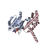
| Unit cell |
|
-Components
| #1: Protein | Mass: 36459.453 Da / Num. of mol.: 1 Fragment: central and catalytic domains of Trf4p (UNP residues 161-481) Mutation: D293A Source method: isolated from a genetically manipulated source Source: (gene. exp.) Gene: PAP2, TRF4, YOL115W, O0716, HRC584 / Production host:  References: UniProt: P53632, Transferases; Transferring phosphorus-containing groups; Nucleotidyltransferases | ||
|---|---|---|---|
| #2: Protein | Mass: 9399.759 Da / Num. of mol.: 1 Fragment: fourth and fifth zinc knuckles of Air2p (UNP residues 118-198) Source method: isolated from a genetically manipulated source Source: (gene. exp.) Gene: AIR2, YDL175C / Production host: | ||
| #3: Chemical |   Mass: 65.409 Da / Num. of mol.: 2 / Source method: obtained synthetically / Formula: Zn Mass: 65.409 Da / Num. of mol.: 2 / Source method: obtained synthetically / Formula: Zn#4: Water | ChemComp-HOH / |  Mass: 18.015 Da / Num. of mol.: 58 / Source method: isolated from a natural source / Formula: H2O Mass: 18.015 Da / Num. of mol.: 58 / Source method: isolated from a natural source / Formula: H2O |
-Experimental details
-Experiment
| Experiment | Method: X-RAY DIFFRACTION / Number of used crystals: 1 |
|---|
- Sample preparation
| Crystal | Density Matthews: 2.8 Å3/Da / Density % sol: 56.13 % |
|---|---|
| Crystal grow | Temperature: 277 K / Method: vapor diffusion, hanging drop / pH: 5.8 Details: 100 mM sodium citrate at pH 5.8, 200 mM sodium acetate, 11% PEG 4000, VAPOR DIFFUSION, HANGING DROP, temperature 277K |
-Data collection
| Diffraction | Mean temperature: 110 K | |||||||||||||||
|---|---|---|---|---|---|---|---|---|---|---|---|---|---|---|---|---|
| Diffraction source | Source: SYNCHROTRON / Site: APS  / Beamline: 24-ID-C / Wavelength: 1.28215 Å / Beamline: 24-ID-C / Wavelength: 1.28215 Å | |||||||||||||||
| Detector | Type: ADSC QUANTUM 315 / Detector: CCD / Date: Apr 19, 2009 | |||||||||||||||
| Radiation | Monochromator: cryogenically cooled Si double crystal (111) monochromator Protocol: SAD / Monochromatic (M) / Laue (L): M / Scattering type: x-ray | |||||||||||||||
| Radiation wavelength | Wavelength: 1.28215 Å / Relative weight: 1 | |||||||||||||||
| Reflection | Resolution: 2.6→25 Å / Num. all: 16163 / Num. obs: 16163 / % possible obs: 100 % / Observed criterion σ(F): 0 / Observed criterion σ(I): -3 / Redundancy: 10.6 % / Rmerge(I) obs: 0.077 | |||||||||||||||
| Reflection shell |
|
- Processing
| Software |
| |||||||||||||||||||||||||||||||||||||||||||||||||||||||||||||||||||||||||||||||||||||||||||||||||||||||||||||||||||||||||||||
|---|---|---|---|---|---|---|---|---|---|---|---|---|---|---|---|---|---|---|---|---|---|---|---|---|---|---|---|---|---|---|---|---|---|---|---|---|---|---|---|---|---|---|---|---|---|---|---|---|---|---|---|---|---|---|---|---|---|---|---|---|---|---|---|---|---|---|---|---|---|---|---|---|---|---|---|---|---|---|---|---|---|---|---|---|---|---|---|---|---|---|---|---|---|---|---|---|---|---|---|---|---|---|---|---|---|---|---|---|---|---|---|---|---|---|---|---|---|---|---|---|---|---|---|---|---|---|
| Refinement | Method to determine structure: SAD / Resolution: 2.7007→24.656 Å / SU ML: 0.37 / σ(F): 0.14 / Phase error: 27.04 / Stereochemistry target values: ML
| |||||||||||||||||||||||||||||||||||||||||||||||||||||||||||||||||||||||||||||||||||||||||||||||||||||||||||||||||||||||||||||
| Solvent computation | Shrinkage radii: 0.9 Å / VDW probe radii: 1.11 Å / Solvent model: FLAT BULK SOLVENT MODEL / Bsol: 39.544 Å2 / ksol: 0.315 e/Å3 | |||||||||||||||||||||||||||||||||||||||||||||||||||||||||||||||||||||||||||||||||||||||||||||||||||||||||||||||||||||||||||||
| Displacement parameters |
| |||||||||||||||||||||||||||||||||||||||||||||||||||||||||||||||||||||||||||||||||||||||||||||||||||||||||||||||||||||||||||||
| Refinement step | Cycle: LAST / Resolution: 2.7007→24.656 Å
| |||||||||||||||||||||||||||||||||||||||||||||||||||||||||||||||||||||||||||||||||||||||||||||||||||||||||||||||||||||||||||||
| Refine LS restraints |
| |||||||||||||||||||||||||||||||||||||||||||||||||||||||||||||||||||||||||||||||||||||||||||||||||||||||||||||||||||||||||||||
| LS refinement shell |
| |||||||||||||||||||||||||||||||||||||||||||||||||||||||||||||||||||||||||||||||||||||||||||||||||||||||||||||||||||||||||||||
| Refinement TLS params. | Method: refined / Refine-ID: X-RAY DIFFRACTION
| |||||||||||||||||||||||||||||||||||||||||||||||||||||||||||||||||||||||||||||||||||||||||||||||||||||||||||||||||||||||||||||
| Refinement TLS group |
|
