 Movie
Movie Controller

[English] 日本語
 Yorodumi
- PDB-7l5b: Crystallographic structure of neutralizing antibody 2-15 in compl... -
+ Open data

- Basic information
| Entry | Database: PDB / ID: 7l5b | ||||||
|---|---|---|---|---|---|---|---|
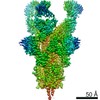
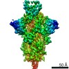
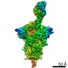
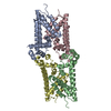
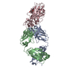
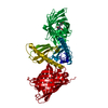
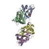
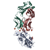
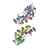
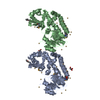
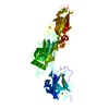
| Title | Crystallographic structure of neutralizing antibody 2-15 in complex with SARS-CoV-2 spike receptor-binding Domain (RBD). | ||||||
 Components Components |
| ||||||
 Keywords Keywords | VIRAL PROTEIN/Immune System / Spike protein S1 / receptor binding protein SARS COV-2 / VIRAL PROTEIN / VIRAL PROTEIN-Immune System complex | ||||||
| Function / homology |  Function and homology information Function and homology informationsymbiont-mediated disruption of host tissue / Maturation of spike protein / Translation of Structural Proteins / Virion Assembly and Release / host cell surface / host extracellular region / symbiont-mediated-mediated suppression of host tetherin activity / Induction of Cell-Cell Fusion / structural constituent of virion / positive regulation of viral entry into host cell ...symbiont-mediated disruption of host tissue / Maturation of spike protein / Translation of Structural Proteins / Virion Assembly and Release / host cell surface / host extracellular region / symbiont-mediated-mediated suppression of host tetherin activity / Induction of Cell-Cell Fusion / structural constituent of virion / positive regulation of viral entry into host cell / membrane fusion / host cell endoplasmic reticulum-Golgi intermediate compartment membrane / Attachment and Entry / entry receptor-mediated virion attachment to host cell / receptor-mediated virion attachment to host cell / host cell surface receptor binding / symbiont-mediated suppression of host innate immune response / endocytosis involved in viral entry into host cell / receptor ligand activity / fusion of virus membrane with host plasma membrane / fusion of virus membrane with host endosome membrane / viral envelope / symbiont entry into host cell / virion attachment to host cell / host cell plasma membrane / SARS-CoV-2 activates/modulates innate and adaptive immune responses / virion membrane / membrane / identical protein binding / plasma membrane Similarity search - Function | ||||||
| Biological species |   Severe acute respiratory syndrome coronavirus 2 Severe acute respiratory syndrome coronavirus 2 Homo sapiens (human) Homo sapiens (human) | ||||||
| Method |  X-RAY DIFFRACTION / SYNCHROTRON / MOLECULAR REPLACEMENT / Resolution: 3.18 Å X-RAY DIFFRACTION / SYNCHROTRON / MOLECULAR REPLACEMENT / Resolution: 3.18 Å | ||||||
 Authors Authors | Reddem, E.R. / Shapiro, L. | ||||||
| Funding support |  China, 1items China, 1items
| ||||||
 Citation Citation | Journal: Cell Rep / Year: 2021 Title: Modular basis for potent SARS-CoV-2 neutralization by a prevalent VH1-2-derived antibody class. Authors: Micah Rapp / Yicheng Guo / Eswar R Reddem / Jian Yu / Lihong Liu / Pengfei Wang / Gabriele Cerutti / Phinikoula Katsamba / Jude S Bimela / Fabiana A Bahna / Seetha M Mannepalli / Baoshan ...Authors: Micah Rapp / Yicheng Guo / Eswar R Reddem / Jian Yu / Lihong Liu / Pengfei Wang / Gabriele Cerutti / Phinikoula Katsamba / Jude S Bimela / Fabiana A Bahna / Seetha M Mannepalli / Baoshan Zhang / Peter D Kwong / Yaoxing Huang / David D Ho / Lawrence Shapiro / Zizhang Sheng /  Abstract: Antibodies with heavy chains that derive from the VH1-2 gene constitute some of the most potent severe acute respiratory syndrome coronavirus 2 (SARS-CoV-2)-neutralizing antibodies yet identified. To ...Antibodies with heavy chains that derive from the VH1-2 gene constitute some of the most potent severe acute respiratory syndrome coronavirus 2 (SARS-CoV-2)-neutralizing antibodies yet identified. To provide insight into whether these genetic similarities inform common modes of recognition, we determine the structures of the SARS-CoV-2 spike in complex with three VH1-2-derived antibodies: 2-15, 2-43, and H4. All three use VH1-2-encoded motifs to recognize the receptor-binding domain (RBD), with heavy-chain N53I-enhancing binding and light-chain tyrosines recognizing F486. Despite these similarities, class members bind both RBD-up and -down conformations of the spike, with a subset of antibodies using elongated CDRH3s to recognize glycan N343 on a neighboring RBD-a quaternary interaction accommodated by an increase in RBD separation of up to 12 Å. The VH1-2 antibody class, thus, uses modular recognition encoded by modular genetic elements to effect potent neutralization, with the VH-gene component specifying recognition of RBD and the CDRH3 component specifying quaternary interactions. #1: Journal: Biorxiv / Year: 2021Title: Modular basis for potent SARS-CoV-2 neutralization by a prevalent VH1-2-derived antibody class Authors: Rapp, M. / Guo, Y. / Reddem, E.R. / Liu, L. / Wang, P. / Yu, J. / Cerutti, G. / Bimela, J. / Bahna, F. / Mannepalli, S. / Zhang, B. / Kwong, P.D. / Ho, D.D. / Shapiro, L. / Sheng, Z. | ||||||
| History |
|
- Structure visualization
| Structure viewer | Molecule: MolmilJmol/JSmol |
|---|
- Downloads & links
-Download
| PDBx/mmCIF format | 7l5b.cif.gz | 129.1 KB | Display | PDBx/mmCIF format |
|---|---|---|---|---|
| PDB format | pdb7l5b.ent.gz | 98.2 KB | Display | PDB format |
| PDBx/mmJSON format | 7l5b.json.gz | Tree view | PDBx/mmJSON format | |
| Others |  Other downloads Other downloads |
-Validation report
| Arichive directory | https://data.pdbj.org/pub/pdb/validation_reports/l5/7l5bftp://data.pdbj.org/pub/pdb/validation_reports/l5/7l5b | HTTPS FTP |
|---|
-Related structure data
| Related structure data | 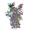 7l56C 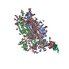 7l57C  7l58C 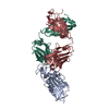 7bz5S S: Starting model for refinement C: citing same article ( |
|---|---|
| Similar structure data |


-Links
 PDBj
PDBj


- Assembly
| Deposited unit | 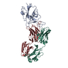
| ||||||||
|---|---|---|---|---|---|---|---|---|---|
| 1 |
| ||||||||
| Unit cell |
|
-Components
| #1: Protein | Mass: 26898.271 Da / Num. of mol.: 1 Source method: isolated from a genetically manipulated source Source: (gene. exp.) Severe acute respiratory syndrome coronavirus 2Gene: S, 2 / Production host: Homo sapiens (human) / References: UniProt: P0DTC2 |
|---|---|
| #2: Antibody | Mass: 24333.307 Da / Num. of mol.: 1 Source method: isolated from a genetically manipulated source Source: (gene. exp.) Homo sapiens (human) / Production host: Homo sapiens (human) |
| #3: Antibody | Mass: 22755.125 Da / Num. of mol.: 1 Source method: isolated from a genetically manipulated source Source: (gene. exp.) Homo sapiens (human) / Production host: Homo sapiens (human) |
| Has protein modification | Y |
-Experimental details
-Experiment
| Experiment | Method: X-RAY DIFFRACTION / Number of used crystals: 1 |
|---|
- Sample preparation
| Crystal | Density Matthews: 4.43 Å3/Da / Density % sol: 72.26 % |
|---|---|
| Crystal grow | Temperature: 298 K / Method: vapor diffusion, sitting drop / pH: 7.5 / Details: 0.1 M Hepes, 70 % MPD |
-Data collection
| Diffraction | Mean temperature: 100 K / Serial crystal experiment: N |
|---|---|
| Diffraction source | Source: SYNCHROTRON / Site: APS / Beamline: 24-ID-C / Wavelength: 0.97918 Å |
| Detector | Type: DECTRIS EIGER2 X 16M / Detector: PIXEL / Date: Oct 14, 2020 |
| Radiation | Protocol: SINGLE WAVELENGTH / Monochromatic (M) / Laue (L): M / Scattering type: x-ray |
| Radiation wavelength | Wavelength: 0.97918 Å / Relative weight: 1 |
| Reflection | Resolution: 3.18→102.49 Å / Num. obs: 27227 / % possible obs: 99.8 % / Redundancy: 4.5 % / CC1/2: 0.951 / Rmerge(I) obs: 0.111 / Net I/σ(I): 1.7 |
| Reflection shell | Resolution: 3.18→3.88 Å / Rmerge(I) obs: 0.11 / Num. unique obs: 4388 / CC1/2: 0.992 |
- Processing
| Software |
| ||||||||||||||||||||||||||||||||||||||||||||||||||||||||||||||||||||||||||||||||||||||||||||||||||||||||||||||||
|---|---|---|---|---|---|---|---|---|---|---|---|---|---|---|---|---|---|---|---|---|---|---|---|---|---|---|---|---|---|---|---|---|---|---|---|---|---|---|---|---|---|---|---|---|---|---|---|---|---|---|---|---|---|---|---|---|---|---|---|---|---|---|---|---|---|---|---|---|---|---|---|---|---|---|---|---|---|---|---|---|---|---|---|---|---|---|---|---|---|---|---|---|---|---|---|---|---|---|---|---|---|---|---|---|---|---|---|---|---|---|---|---|---|
| Refinement | Method to determine structure: MOLECULAR REPLACEMENT Starting model: 7bz5 Resolution: 3.18→102.49 Å / SU ML: 0.31 / Cross valid method: FREE R-VALUE / σ(F): 0 / Phase error: 21.7 / Stereochemistry target values: ML
| ||||||||||||||||||||||||||||||||||||||||||||||||||||||||||||||||||||||||||||||||||||||||||||||||||||||||||||||||
| Solvent computation | Shrinkage radii: 0.9 Å / VDW probe radii: 1.11 Å / Solvent model: FLAT BULK SOLVENT MODEL | ||||||||||||||||||||||||||||||||||||||||||||||||||||||||||||||||||||||||||||||||||||||||||||||||||||||||||||||||
| Refinement step | Cycle: LAST / Resolution: 3.18→102.49 Å
| ||||||||||||||||||||||||||||||||||||||||||||||||||||||||||||||||||||||||||||||||||||||||||||||||||||||||||||||||
| Refine LS restraints |
| ||||||||||||||||||||||||||||||||||||||||||||||||||||||||||||||||||||||||||||||||||||||||||||||||||||||||||||||||
| LS refinement shell |
|
